


 64 / 67

CARDIOVASCULAR JOURNAL OF AFRICA • Vol 24, No 2, March 2013
e14
AFRICA
central vein catheter over the next five minutes, with subsequent
stabilisation of his BP.
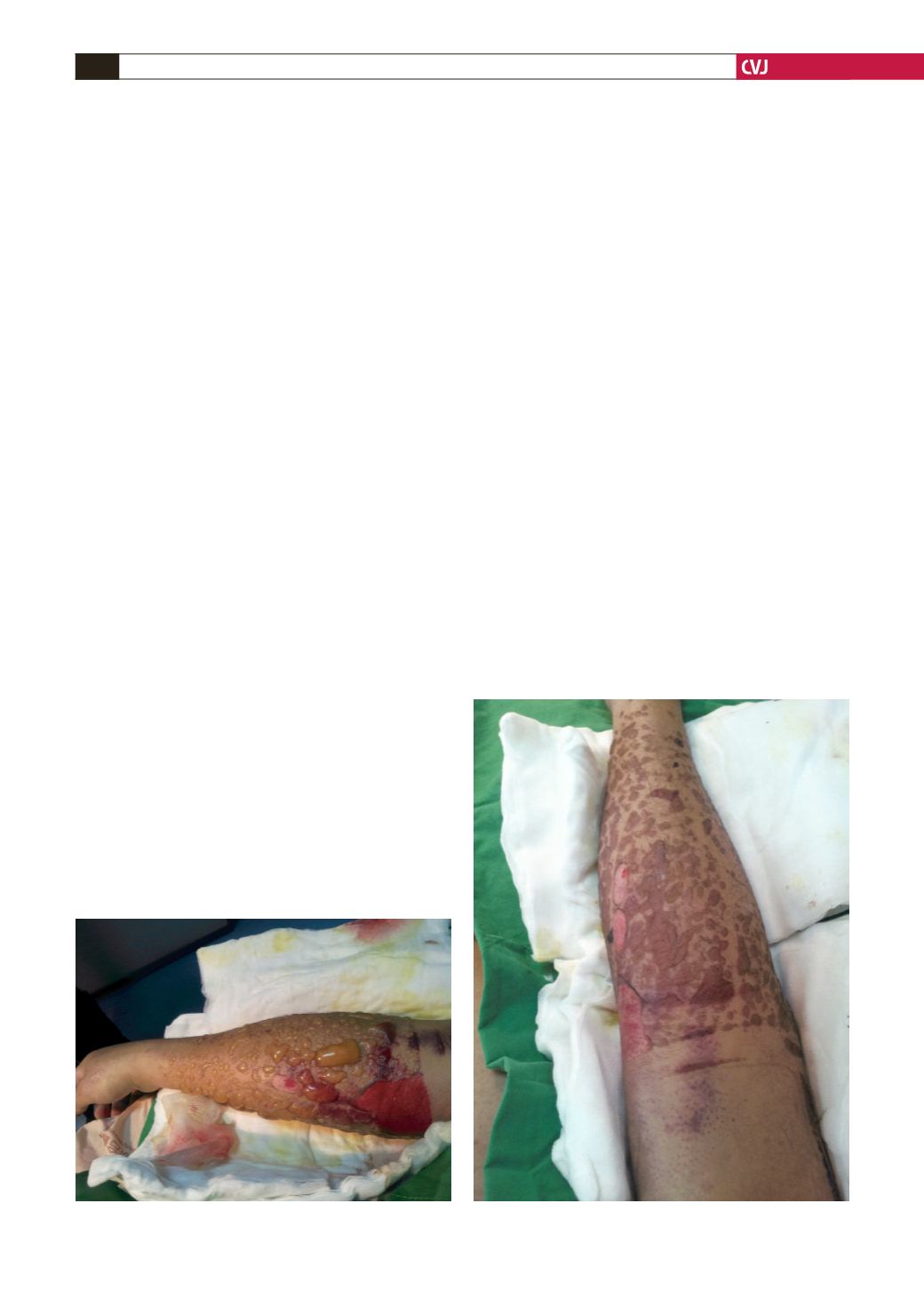
After the operation, we noted the localised bullous eruptions
on the right forearm (Fig. 1). Further examination revealed red,
swollen blisters over the right forearm (Fig. 2). This condition
had not been noticed earlier. Digital capillary refill was noted
to be delayed compared to the contralateral extremity. The
NIBP cuff and right antecubital catheter were removed and the
cardiovascular team was alerted. Doppler monitoring detected
diminished right radial and ulnar pulses.
Extravasation of the fluids infused under pressure was the
apparent aetiology. Initially, conservative treatment was chosen
with needle aspiration of the fluid in the localised eruptions. We
then sent the patient to the intensive care unit with protective
gauze covering the right forearm. The right upper extremity
was closely monitored during the initial postoperative period.
Neurological and vascular functions remained intact.
Seven days after the operation, the forearm oedema gradually
decreased and the wound was clean. He underwent heart
transplantation one month later. Follow up at six months
identified no long-term sequelae of his upper extremity injury.
Discussion
Peripheral intravenous infusion of agents is a daily routine in
hospitals. Extravasation injury is defined as damage caused
by leakage of fluid from a vein into the surrounding tissue
spaces during intravenous infusion. Extravasation of intravenous
infusions is one of the iatrogenic complications frequently
encountered in hospitals.
For central venous catheters, extravasation is less frequent but
potentially more dangerous because the anatomical structures
escape attention. Depending on the insertion depth, the extravasal
position of the proximal port can occur when the catheter is
inadvertently withdrawn just a few centimetres.
1
Extravasation
may also go unnoticed in peripheral lines when the area is covered
by drapes during surgery. This has led clinicians to underestimate
the potentially serious consequences of extravasation.
Common sites of injury are the dorsum of the hand (extension
crease to the metacarpophalangeal joint) and the antecubital
fossa, where there is little soft-tissue coverage.
3,4
Venous
extravasation is caused by escape of the needle or cannula tip
from the vessel lumen through accidental pull out, penetration
of the counter vessel wall, or injection of large volumes or at a
fast rate through the infusion pump with the needle still inside
the vein.
2,3
However, in most cases, the tip of the cannula remains
in the lumen and extravasation is through the hole made by the
cannula or needle entering the vein.
3,4
Extravasation is relatively more common in elderly or
cachetic patients, whose veins are more fragile, and the puncture
hole from the cannula is easier to enlarge, which may cause a
leak. Our patient was a typical example. The vascular supply
to the skin has been described as segmental perforator systems.
Extravasation of fluid stretches the vessels, leading to partial
venous occlusion, followed by arterial occlusion.
4
The resulting
increase in intraluminal pressure leads to leakage of fluid from
the puncture site.
3
In the peri-operative period, the mechanism of tissue necrosis
can include solution cytotoxicity, osmolality, vasoconstrictor
effects, infusion pressure, regional anatomical peculiarities, and
other factors.
1,4
If this continues for an extended period of time,
cellular death and skin breakdown follow.
In our case, blistering was a sign of serious skin injury,
possibly resulting from oedema. The depth and extent of tissue
injury depend on factors such as the volume of fluid extravasated,
composition of the fluid, location of the leak, passage of time
before the accident is discovered, and the measures taken after
discovery of the incident.
Given this situation, intra-operative attention and on-going
patient assessment by the anaesthesiologist is important. In
our case, a swollen forearm and diminished radial pulse were
the only findings that prompted further investigation. Prompt
Fig. 2. Red, swollen blisters over the right forearm.
Fig. 1. Localised bullous eruptions on the right forearm.