


 50 / 78

CARDIOVASCULAR JOURNAL OF AFRICA • Volume 26, No 4, July/August 2015
196
AFRICA
First Melody
®
valve implantations in Africa
DG Buys, C Greig, SC Brown
Abstract
Congenital heart lesions involving the right ventricular
outflow tract (RVOT) are a common problem in paediatric
cardiology. These patients need multiple surgical interven-
tions in the form of valved conduits over a lifetime. Surgical
re-valvulation was the standard treatment option until the
introduction of percutaneous pulmonary valves over a decade
ago. These valves can be used to prolong the lifespan of
conduits and reduce the number of re-operations. The
Melody
®
valve (Medtronic, Minneapolis, MN, USA) was
introduced as the first dedicated percutaneous pulmonary
valve. Percutaneous pulmonary valves can be implanted
successfully and have the advantage of short hospitalisations.
We describe the first three Melody
®
valve implantations in
Africa.
Keywords:
Melody
®
valve, Africa, percutaneous valve, implanta-
tion
Submitted 12/12/13, accepted 11/1/15
Cardiovasc J Afr
2015;
26
: 196–199
www.cvja.co.zaDOI: 10.5830/CVJA-2015-007
Right ventricle-to-pulmonary artery (RV–PA) conduit failure
is a vexing problem in post-operative congenital cardiac
lesions involving the right ventricular outflow tract (RVOT).
Typical lesions include tetralogy of Fallot, pulmonary atresia,
truncus arteriosus, and others. These lesions often require
early intervention and multiple RVOT revisions. Surgical
re-interventions may result in prolonged hospital stay with
increased morbidity and mortality rates.
1,2
Due to the invasive
nature of surgery, some patients with RVOT dysfunction are
managed for years before surgical re-valvulation is considered.
The first percutaneous pulmonary valve was implanted in
the year 2000, and led to the development of the Melody
®
valve
(Medtronic, Minneapolis, MN, USA).
3-5
The Melody
®
valve
consists of an 18-mm valve segment, the Contegra
®
modified
bovine jugular vein, sutured into a platinum iridium stent of
34-mm length (Fig. 1). The valve can be crimped down to
6 mm and re-expanded from 18 to 22 mm using the Ensemble
®
transcatheter delivery system (Medtronic, Minneapolis, MN,
USA).
We describe the first three Melody
®
valve implantations in
Africa. These were done at the Universitas Academic Hospital
complex in Bloemfontein, South Africa.
Case report
Only patients meeting standard indications for surgical
re-intervention were evaluated for transcatheter valve
implantation. Extensive work up included: chest radiography,
electrocardiography (ECG), evaluation of exercise capacity,
echocardiography, and high-resolution computed tomographic
angiography (CTA) (Fig. 2). The right ventricle size and function
as well as the severity of pulmonary regurgitation (PR) and/or
pulmonary stenosis (PS) were assessed and quantified. CTA in all
three patients demonstrated favourable coronary artery anatomy
and we proceeded with valve implantation in March 2012.
Case 1
The patient was a 17-year-old male (weight 46.2 kg) with
tetralogy of Fallot. As initial intervention, he had had surgical
correction and a RVOT patch at 18 months of age. This was later
followed by a RV-PA outflow tract reconstruction and insertion
of a 20-mm homograft at age 11 years. He was considered for
percutaneous valve implantation because of exercise intolerance,
right ventricular dysfunction and severe PR.
Department of Paediatric Cardiology, University of the Free
State, and Universitas Hospital, Bloemfontein, South Africa
DG Buys, MMed (Paed), Cert Paed Cardiol,
buysdg@ufs.ac.zaC Greig, MMed (Paed)
SC Brown, MMed (Paed), FCPaed (Cardio)
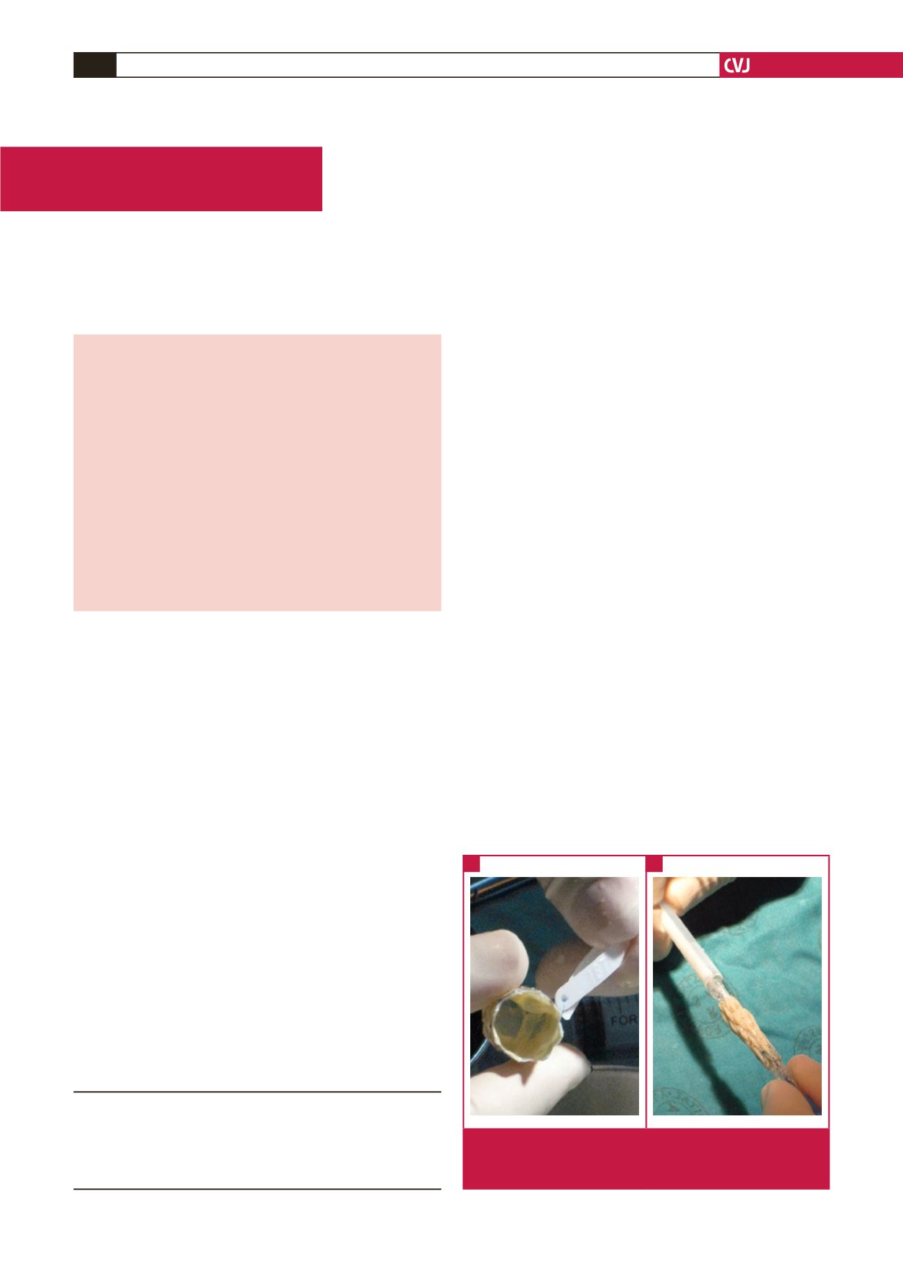
Fig. 1.
A: competent Melody
®
valve with ID tag, B: the valve
crimped on to the Ensemble
®
delivery system pre valve
implantation.
A
B
Case Report