CARDIOVASCULAR JOURNAL OF AFRICA • Vol 23, No 8, September 2012
AFRICA
437
UACR, both in type 1 and type 2 diabetes patients (all
p
<
0.05)
(
Table 4). In addition, lower eGFR and high-density lipoprotein
(
HDL) cholesterol were significantly correlated with higher
RWT among type 2 but not in type 1 diabetes patients. Having
increased RWT was also associated with impaired systolic and
diastolic LV function, including lower myocardial contractility,
measured as scMWS, and delayed early LV diastolic relaxation,
measured as longer IVRT, longer deceleration time and reduced
E/A ratio, both in type 1 and type 2 diabetes patients (all
p
<
0.05) (
Table 4).
When multivariate linear regression analyses were performed,
higher systolic blood pressure, longer IVRT and low scMWS
remained significant covariates of higher RWT both in type 1
and type 2 diabetes patients, irrespective of presence or absence
of LV hypertrophy and also adjusted for CESS. In addition, low
eGFR continued to be an independent covariate of higher RWT
in type 2 diabetes patients. Substituting log UACR for eGFR in
the type 1 diabetes patients’ model did not give any independent
association either (Table 5).
In binary logistic regression analysis, including type of
diabetes, albuminuria, obesity, history of hypertension and HbA
1
c
level, the independent covariates of increased RWT were: type
2
diabetes (OR 2.7, 95% CI: 1.08–7.00), albuminuria (OR 2.2,
95%
CI: 1.01–4.62), obesity (OR 2.6, 95% CI: 1.02–6.58) and
hypertension (OR 2.5, 95% CI: 1.02–5.87), all
p
<
0.05.
A risk score was calculated based on the beta coefficients in
this model: risk score
=
9
x
(
type of diabetes)
+
8
x
(
albuminuria)
+ 9
x
(
obesity) + 9
x
(
hypertension). For each parameter included
in the score, a value of 1 was assigned if the variable was present
TABLE 2. ECHOCARDIOGRAPHIC FINDINGS IN TYPE 1 AND TYPE 2 DIABETES PATIENTS
Unadjusted
Adjusted for age and systolic blood pressure
Echocardiographic finding
Type 1
(
n
=
61)
Type 2
(
n
=
123)
p
-
value
Type 1
(
n
=
61)
Type 2
(
n
=
123)
p
-
value
Interventricular septum in diastole (cm)
0.91
±
0.21
1.27
±
0.31
<
0.001
1.11
±
0.06
1.16
±
0.04
0.573
LV posterior wall in diastole (cm)
0.79
±
0.17
1.06
±
0.25
<
0.001
0.94
±
(.05
0.98
±
0.03
0.622
LV end-diastolic diameter (cm)
4.01
±
0.63
4.21
±
0.58
0.036
4.10
±
0.13
4.16
±
0.08
0.769
Relative wall thickness
0.40
±
0.10
0.52
±
0.19
<
0.001
0.48
±
0.04
0.48
±
0.02
0.938
LV mass/height
2.7
(
g/m
2.7
)
33.0
±
9.6
49.2
±
16.8
<
0.001
40.6
±
3.0
45.1
±
1.8
0.299
Fractional shortening (%)
37
±
5
35
±
6
0.176
36
±
1.3
36
±
0.8
0.940
Stress-corrected fractional shortening (%)
99
±
11
99
±
16
0.942
100
±
3
99
±
2
0.739
Ejection fraction (%)
65
±
7
63
±
8
0.328
63
±
2
64
±
1
0.554
Midwall shortening (%)
16
±
3
13
±
3
<
0.001
14
±
0.7
15
±
0.4
0.875
Stress-corrected midwall shortening (%)
90
±
17
74
±
18
<
0.001
80
±
3.8
81
±
2.4
0.918
Transmitral E/A ratio
1.5
±
0.4
0.9
±
0.3
<
0.001
1.2
±
0.8
1.1
±
0.5
0.226
Deceleration time (ms)
165
±
52
206
±
61
<
0.001
191
±
13
192
±
8
0.954
Isovolumic relaxation time (ms)
62
±
16
81
±
20
<
0.001
78
±
3.8
73
±
2.4
0.378
Early tissue Doppler velocity (E
′
) (
cm/s)
10.3
±
2.3
6.5
±
2.4
<
0.001
8.3
±
0.5
7.5
±
0.3
0.305
E/E
′
ratio
9.5
±
2.4
11.7
±
4.4
<
0.001
11.2
±
0.8
10.8
±
0.5
0.733
TABLE 1. DEMOGRAPHICAND LABORATORY
CHARACTERISTICS OF TYPE 1 AND
TYPE 2 DIABETES PATIENTS
Characteristic
Type 1
(
n
=
61)
Type 2
(
n
=
123)
p
-
value
Age (years)
21.7
±
10.6 55.0
±
9.6
<
0.001
Females,
n
(%)
34 (55)
78 (64)
0.265
Duration of diabetes (years)
8.2
±
4.5 10.7
±
6.3 0.005
Body mass index (kg/m
2
)
20.9
±
4.4 28.4
±
4.7
<
0.001
Obesity,
n
(%)
2 (3.3)
45 (36.6)
<
0.001
Waist circumference (cm)
74
±
12
98
±
11
<
0.001
Systolic blood pressure (mmHg)
117
±
21 147
±
22
<
0.001
Diastolic blood pressure (mmHg)
74
±
14
88
±
11
<
0.001
Hypertension,
n
(%)
11 (17.7)
100 (82.0)
<
0.001
Pulse pressure (mmHg)
43
±
12
59
±
17
<
0.001
Fasting blood glucose (mmol/l)
12.2
±
4.4 10.4
±
4.7 0.015
HbA
1
c
(%)
10.9
±
2.2 9.8
±
2.3 0.003
Total cholesterol (mmol/l)
4.7
±
1.6 5.6
±
1.5 0.001
HDL cholesterol (mmol/l)
1.2
±
0.4 1.2
±
0.3 0.855
LDL cholesterol (mmol/l)
3.2
±
1.3 4.0
±
1.4
<
0.001
Triglycerides (mmol/l)
1.6
±
1.6 1.7
±
1.0 0.617
Serum creatinine (
µ
mol/l)
84
±
70 106
±
77 0.058
eGFR (ml/min/1.73 m
2
)
106
±
47 81
±
24
<
0.001
Low eGFR,
n
(%)
6 (10)
21 (18)
0.268
Albuminuria,
n
(%)
24 (40.0)
39 (33.6)
0.412
Microalbuminuria,
n
(%)
16 (26.7)
33 (28.4)
0.860
Macroalbuminuria,
n
(%)
8 (13.3)
6 (5.2)
0.077
HbA
1
c
=
glycated haemoglobin, HDL
=
high-density lipoprotein, LDL
=
low-density lipoprotein, eGFR
=
estimated glomerular filtration rate.
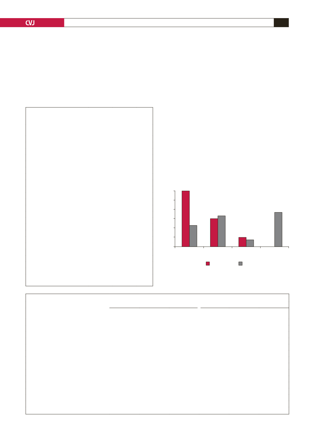
60
50
40
30
20
10
0
Percent
Normal
Concentric
remodelling
Eccentric Concentric
LVH
60
23
30 33
10 7.3
36.7
0
Type 1
Type 2
Fig. 1. LV geometric patterns in type 1 (red bars) and type
2 (
grey bars) diabetes patients. The differences between
normal geometry and concentric LVH were statistically
significant, both
p
<
0.001.


