CARDIOVASCULAR JOURNAL OF AFRICA • Vol 23, No 8, September 2012
AFRICA
453
<
0.05).
In addition, heart-to-body weight ratio, as an index of
heart hypertrophy, was greater in the EXT rats than the sedentary
ones (
p
<
0.05).
Fig. 1 shows a progressive increase in the weight-lifting
ability of the EXT rats. Both the Sed and EXT groups had
similar values for work performed in the first (week 1) of the
protocol. The work performed at the end of weeks 2, 3 and 4
were significantly higher in the EXT rats than the Sed group (
p
<
0.05,
p
<
0.01
and
p
<
0.01,
respectively) and their previous
week’s values (
p
<
0.05,
p
<
0.05
and
p
<
0.01,
respectively).
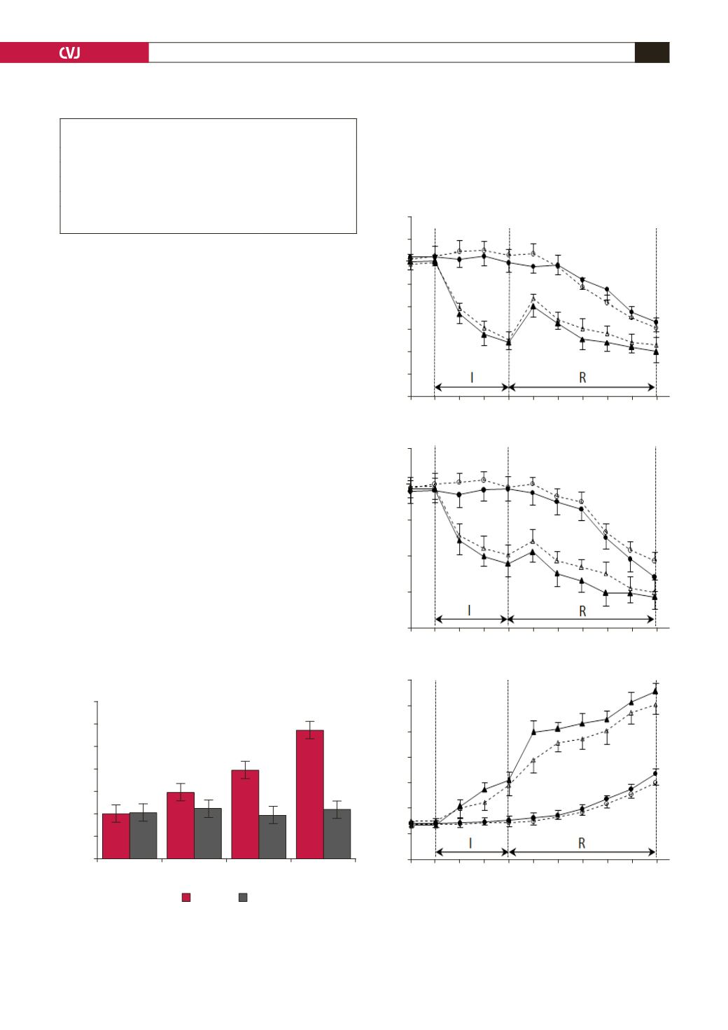
Developed pressure, diastolic pressure and coronary flow
changes during the time-control and ischaemia–reperfusion
periods for the EXT and Sed groups are shown in Fig.
2.
Baseline coronary flow, developed pressure and diastolic
pressure were similar in the two groups. No between-group
differences in developed or diastolic pressure were observed at
any time point in the non-ischaemic time-control measurements.
While diastolic pressure increased and developed pressure and
coronary flow decreased in both the ischaemia and reperfusion
periods (as indices of cardiac damage), there were no statistically
significant differences between the EXT and Sed groups in these
parameters.
Figs 3 and 4 show the size of the infarction and the apoptosis
rate, respectively in the hearts of the EXT and Sed groups.
Resistance exercise training did not significantly change the
infarct size or apoptosis rate.
Discussion
Our previous study showed that 12-week resistance exercise
training preserved the heart against IR-induced injury.
11
Although
there are some reports on the effect of resistance training on
cardiac structure and function, to the best of our knowledge,
this is the first study that has focused on the role of short-term
resistance training in preserving the heart against IR-induced
Fig. 1. Work performed by rats after the end of each week
of resistance exercise training. Values are mean
±
SD (
n
=
20
rats); *
p
<
0.01, **
p
<
0.05
compared with previous
week;
†
p
<
0.01,
††
p
<
0.05
compared with the sedentary
group; Sed: sedentary and EXT: exercise-trained rats.
1.4
1.2
1
0.8
0.6
0.4
0.2
0
1
2
3
4
Work performed (kg
×
m/day)
Weeks
EXT
Sed
**
††
**
†
†
TABLE1. EFFECTS OF RESISTANCE EXERCISE
ON THE RAT MORPHOLOGY
Sed
EXT
Body weight (g)
266
±
13
259
±
11
Heart weight (g)
0.75
±
0.06
0.84
±
0.06**
Body:heart ratio
2.8
±
0.15
3.2
±
0.18**
Values are mean
±
SD (
n
=
10
rats); **
p
<
0.05,
significantly different
from the sedentary group; Sed: sedentary and EXT: exercise-trained rats.
Fig. 2. Haemodynamic indices of the heart during non-
ischaemic time control (
❍
exercised and
●
sedentary
rats;
n
=
6
for each), regional ischaemia (I) and subse-
quent reperfusion (R) (Δ exercised and
▲
sedentary rats;
n
=
12
for trained and
n
=
11
for sedentary animals). A:
diastolic pressure. B: Left ventricular developed pres-
sure (LVDP). C: Coronary flow. Values are mean
±
SD.
20
18
16
14
12
10
8
6
4
120
100
80
60
40
20
60
50
40
30
20
10
0
–10
Base 0 5 15 40 45 60 75 90 105 120
Base 0 5 15 40 45 60 75 90 105 120
Base 0 5 15 40 45 60 75 90 105 120
Coronary flow (ml/min
×
g)
Developed pressure (mmHg)
Diastolic pressure (mmHg)
A
B
C


