CARDIOVASCULAR JOURNAL OF AFRICA • Vol 23, No 8, September 2012
AFRICA
e7
Case Report
An unusual embolic complication of percutaneous
coronary artery intervention and simple percutaneous
treatment
ERKAN ILHAN, ÖZER SOYLU, TOLGA S GÜVENÇ, YIĞIT ÇANGA, MEHMET ERGELEN
Abstract
Emboli are among the most feared complications of interven-
tional cardiology. Although surgery is needed in most cases
for the removal of peripheric foreign body emboli, some may
be extracted by percutaneous intervention.We present a case
of retrieval of a femoral sheath fragment via contralateral
femoral access, wiring of the sheath fragment, and retrieval
with an ‘anchoring balloon’ system.
Keywords:
percutaneous coronary intervention, femoral sheath,
peripheric embolus
Submitted 18/3/10, accepted 19/3/12
Cardiovasc J Afr
2012;
23
:
e7–e8
DOI: 10.5830/CVJA-2012-030
Case report
A 59-year-old male with a history of hypertension and diabetes
mellitus was admitted to our emergency department with
non-ST-segment elevation myocardial infarction. After medical
treatment and stabilisation of the patient, coronary angiography
was performed from the right femoral artery with a 6F sheath
(
Sentia, Ayra Medical, Turkey).
Drug-eluting stent implantation to the critical left anterior
descending coronary artery lesion was attempted. Unfortunately
the procedure was aborted because of unsuccessful percutaneous
coronary intervention (PCI), and bypass surgery was planned.
Since intravenous heparin had been used during PCI, removal of
the sheath was planned after six hours.
During removal of the sheath, we noticed that the back-bleed
valve and side-arm connector of the sheath had detached from the
shaft. After removal of the valve and side arm, homeostasis was
obtained with manual pressure and the patient was transferred to
the catheter laboratory to visualise the exact position of the shaft.
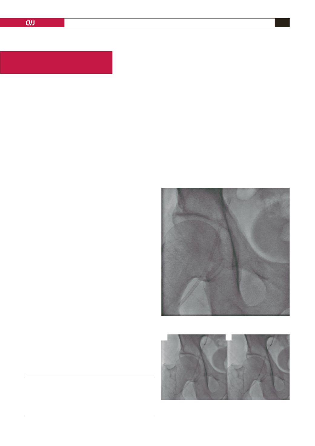
Under fluoroscopy, the shaft was visualised in the right
femoral artery (Fig. 1). Percutaneous removal of the shaft from
the contralateral femoral artery was planed. After insertion
of a 7F sheath (St Jude Medical, Minnetonka, USA) into the
left femoral artery, a 7F left Judkins guiding catheter (Boston
Scientific, Mexico) was advanced into the right external iliac
artery.
When the shaft and the catheter were adjacent, a 0.014-inch
guidewire was passed through both lumens (Fig. 2A). A 2.5
×
Dr Siyami Ersk Cardiovascular and Thoracic Surgery
Training and Research Hospital, Istanbul, Turkey
ERKAN ILHAN, MD,
ÖZER SOYLU, MD
TOLGA S GÜVENÇ, MD
YIĞIT ÇANGA, MD
MEHMET ERGELEN, MD
Fig 1. Shaft of the sheath in the right femoral artery, seen
under fluoroscopy.
A
B
Fig. 2. A 0.014-inch guidewire passed through both
lumens (A) and a 2.5
×
15-
mm angioplasty balloon was
inflated within the shaft.


