CARDIOVASCULAR JOURNAL OF AFRICA • Vol 23, No 8, September 2012
AFRICA
e13
Case Report
Single coronary artery from the right coronary sinus
with proximal origin of the left anterior descending
coronary artery and left circumflex as distal continuation
of the right coronary artery: a rare variant
VIJAYAKUMAR SUBBAN, SUMA M VICTOR, MULLASARI S AJIT, LATCHUMANADHAS KALIDOSS
Abstract
A single coronary artery is a rare coronary anomaly. A
68-
year-old male underwent coronary angiography for recent
inferior wall myocardial infarction. It revealed a common
coronary trunk arising from the right sinus of Valsalva and
bifurcated into the right coronary artery (RCA) and ante-
rior descending coronary arteries. The RCA, after its usual
distribution in the right atrioventricular groove, continued as
the left circumflex artery in the left atrioventricular groove.
There were significant stenoses in the mid and distal RCA,
which were treated percutaneously.
Keywords:
single coronary artery, left anterior descending
artery, left circumflex artery, right coronary artery, percutaneous
coronary intervention
Submitted 31/8/11, accepted 11/4/12
Cardiovasc J Afr
2012;
23
:
e13–e14
DOI: 10.5830/CVJA-2012-034
Coronary artery anomalies are found in 0.6 to 1.5% of coronary
angiograms and are usually incidental findings.
1
Here we
describe a patient with an unusual variant of a single coronary
artery (SCA), who underwent successful percutaneous coronary
intervention.
Case report
A diabetic 65-year-old gentleman with dyslipidaemia presented
to us with a history of recent myocardial infarction. Cardiac
examination was normal and a baseline electrocardiogram
showed QS complexes and T-wave inversions in the inferior
leads.
The echocardiogram revealed wall motion abnormalities
in the right coronary artery (RCA) distribution. On invasive
angiogram, a diminutive coronary artery originated from the left
sinus of Valsalva and followed the course of the first diagonal
branch (D) of the left anterior descending coronary artery (LAD)
(
Fig. 1A). A common coronary trunk was seen to arise from the
right sinus of Valsalva, which bifurcated into the LAD and RCA
immediately distal to the origin (Fig. 1B, C). The LAD followed
a septal course beneath the right ventricular infundibulum into
the anterior interventricular groove.
The RCA continued in the right atrio-ventricular groove
(
AVG) to produce a small posterior descending artery at the crux.
Thereafter, it continued in the left AVG as the left circumflex
Department of Cardiology, Institute of Cardio-Vascular
Diseases, Madras Medical Mission, Chennai, Tamil nadu,
India
VIJAYAKUMAR SUBBAN, MD, DM,
SUMA M VICTOR, DNB (Med), DNB (Cardiol)
MULLASARI S AJIT, MD, DM
LATCHUMANADHAS KALIDOSS, MD, DM
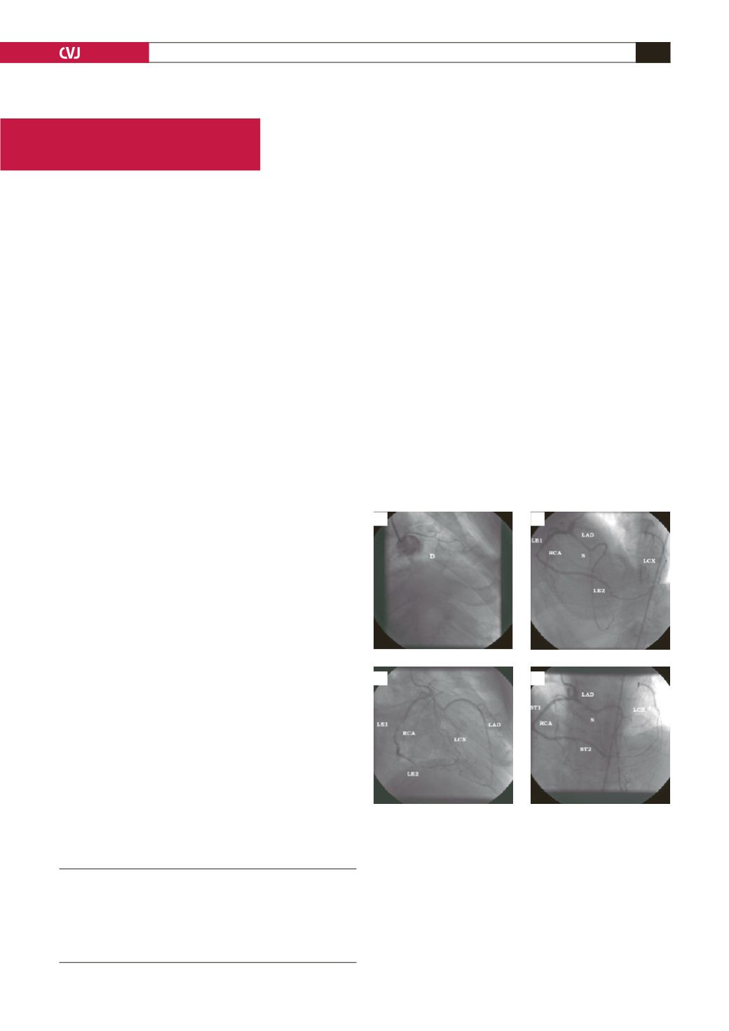
Fig. 1. A. Selective left coronary angiogram showing
a diminutive artery in the diagonal (D) distribution. B.
Selective angiogram of the right-sided common coro-
nary trunk in the left anterior oblique view, showing
early branching into the right coronary artery (RCA) and
left anterior descending coronary artery. The distal RCA
continues as the left circumflex artery. There are stenotic
lesions in the mid and distal RCA. C. Selective angiogram
of the right-sided common coronary trunk in the right
anterior oblique view showing similar features. D. Post-
intervention angiogram in the cranial antero-posterior
view showing the excellent final result. LAD, left anterior
descending coronary artery; LCX, left circumflex artery;
ST, stent; S, septal branch; LE, lesion.
A
C
B
D


