CARDIOVASCULAR JOURNAL OF AFRICA • Vol 23, No 8, September 2012
AFRICA
e9
Case Report
Anomalous left coronary artery arising from the
pulmonary artery
M DURAND, ET NGUYEN, AM CREAN
Abstract
A 24-year-old female presented to her general practitioner
with shortness of breath. She was referred for an echocar-
diogram, which demonstrated features suggestive of a right
coronary artery fistula, and referred to our institute. We
performed a contrast-enhanced, prospectively triggered
cardiac CT angiogram, which demonstrated the primary
and secondary features of anomalous left coronary artery
arising from the pulmonary artery (ALCAPA), also known
as the Bland–White–Garland syndrome, a rare congenital
abnormality of the origin of the left main coronary artery.
Keywords:
anomalous left coronary artery arising from the
pulmonary artery, ALCAPA, congenital coronary abnormality
Submitted 6/11/11, accepted 19/3/12
Cardiovasc J
Afr 2012;
23
:
e9–e10
DOI: 10.5830/CVJA-2012-031
A 24-year-old female presented to her general practitioner with
shortness of breath. She was referred for an echocardiogram,
which demonstrated features suggestive of a right coronary
artery fistula, and she was referred to our institute. We
performed a contrast-enhanced, prospectively triggered cardiac
CT angiogram, using the low-dose 100-kVp technique. Using a
320
multi-detector row scanner (Aquilion One, Toshiba Medical
Systems, Japan), a volumetric acquisition in late diastole was
performed in a single heartbeat.
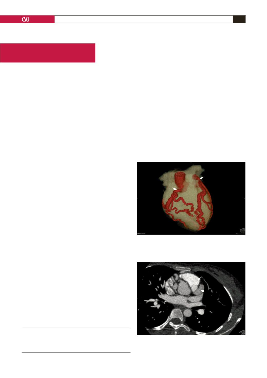
The three-dimensional volume-rendered virtual image (Fig.
1)
and reformatted maximum-intensity projection image (Fig.
2)
demonstrate the left main coronary artery arising from the
left infero-lateral aspect of the main pulmonary artery. The left
main coronary artery extends towards the interventricular groove
and branch into the left anterior descending and left circumflex
coronary arteries. The right coronary artery (RCA) had the
conventional origin from the right coronary sinus of valsalva.
All the coronary arteries were significantly dilated with
multiple intercoronary (Fig. 3) and septal (Fig. 4) collateral
vessels, as seen on the maximum-intensity projection images.
The left ventricle was significantly dilated (not shown) as a result
of the long-term steal of blood from the ascending aorta via the
RCA, to the septal collaterals, into the anomalous left coronary
system and then to the pulmonary bed and left ventricle.
The cardiac CT scan demonstrates the primary and secondary
features of anomalous left coronary artery arising from the
pulmonary artery (ALCAPA), also known as the Bland–White–
Garland syndrome, a rare congenital abnormality of the origin
of the left main coronary artery. This abnormality results in a
coronary steal phenomenon, with right-sided coronary blood
Department of Medical Imaging, University Health Network,
University of Toronto, Canada
M DURAND, MB ChB, FCRad (Diag),
ET NGUYEN, MD, FRCPC
AM CREAN, MD, MRCP, FRCR
Fig. 1. Thee-dimensional volume-rendered image demon-
strating the left main coronary artery (LM) arising from
the main pulmonary artery (MPA). The right coronary
artery (RCA) has a conventional origin from the aorta
(
Ao).
Fig. 2. Reformatted maximum-intensity projection image
in the axial plane demonstrating the left main coronary
artery (LM) arising from the infero-lateral aspect of the
main pulmonary artery (MPA).
LM
MPA
RCA
Ao
LM
MPA
Ao


