


 56 / 67

CARDIOVASCULAR JOURNAL OF AFRICA • Volume 26, No 2, March/April 2015
e2
AFRICA
Following completion of the consultation in the relevant
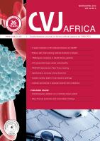
departments, the patient was taken to surgery. The left leg was
operated on first (Fig. 1). Using local anaesthesia, a sheath
through which a radiofrequency ablation catheter (ClosureFast)
®
would move was placed 48 cm from the left lower knee. The
ablation catheter was inserted 2 cm distal to the sapheno-femoral
junction so as to keep the superficial epigastric vein open.
Tumescent anaesthesia was administered throughout the vein
trace to be ablated.
A section of 48 cm of the saphenous vein was ablated
by applying equal power (40 W) to each centimeter. The
ablation procedure was administered three times for each 7-cm
segment. Following the procedure, it was confirmed by Duplex
ultrasonography that the ablated saphenous vein segment
was obliterated, and colour mode of Duplex ultrasonography
showed no flow. In addition, two large excisions of varicose veins
were conducted with phlebectomy from the left lower knee. The
patient was taken to the clinic in tight bandages.
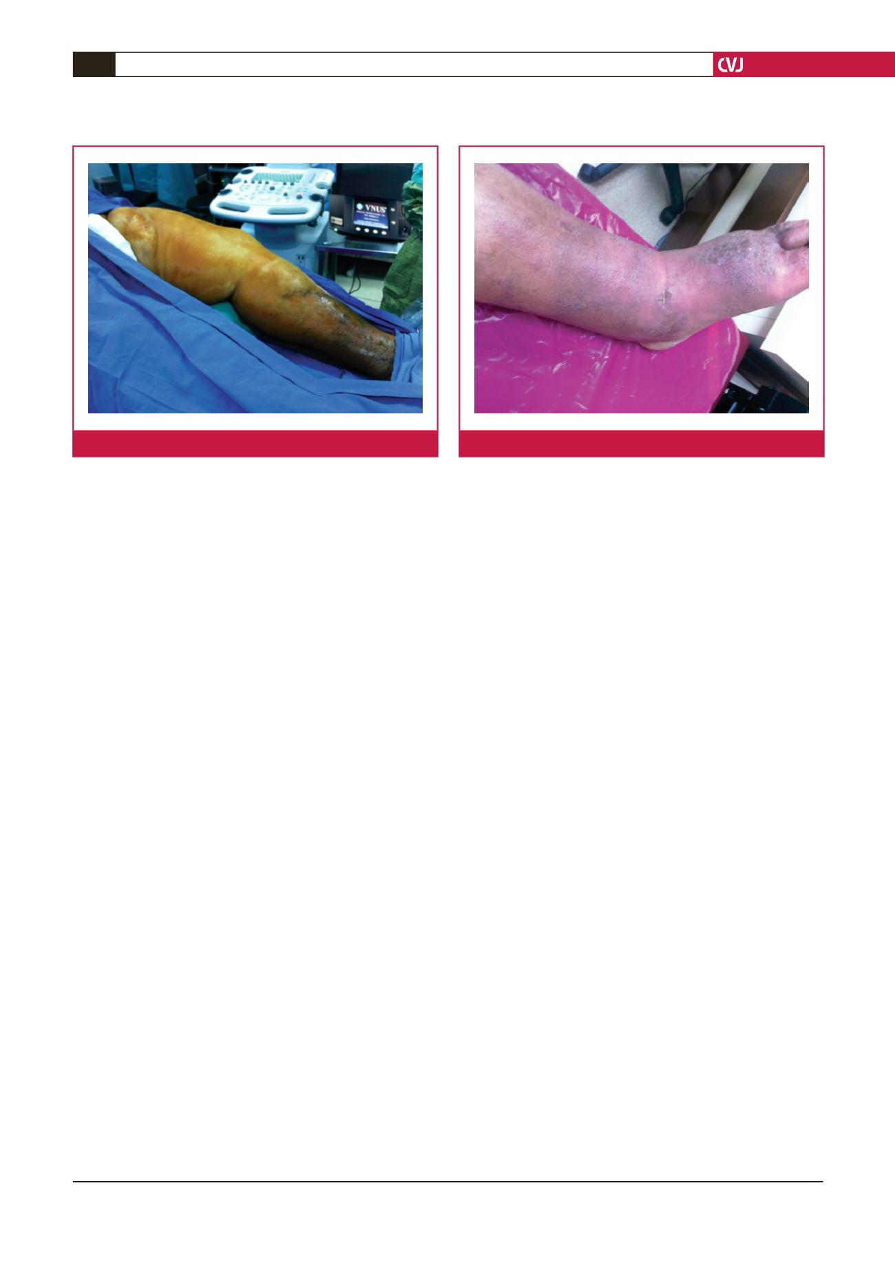
About 21 days later, the right leg was operated on, with
intervention of the saphenous vein in the right lower knee. A
segment of 44 cm of the saphenous vein was ablated and it
was observed to be obliterated following the procedure. Duplex
ultrasonography was done in the first week and first, third and
sixth months after saphenous vein closure and the venous ulcer
was in remission (Fig. 2).
Discussion
Endovenous ablation techniques have been commonly used in
recent years and have started to replace venous stripping, which
was standard in surgery. High patient comfort has promoted
the development of such techniques. Less pain, ecchymosis and
haematoma have increased the usage of these techniques. Such
techniques are being safely used on patients with a high number
of accompanying co-morbid diseases, as in our case, and they
facilitate returning to active life by increasing patient comfort
and allowing them to return to work more quickly.
Radiofrequency ablation procedure produces results
demonstrating that it is better, more comfortable and safer
5
than procedures such as standard surgery and foam therapy.
Procedures conducted with a radiofrequency catheter have
proven to be more successful in patients of all ages.
6
Conclusion
In this context, we believe that radiofrequency ablation, which
can be implemented even for standard surgery cases, is a safe
method for highly varicose and co-morbid patients.
References
1.
Podnos YD, Williams RA, Tessier DJ. Chronic venous in-sufficiency.
Available at:
http://www.emedicine.com/med/topic2760.htm.Accessed
October 25, 2005.
2.
Doughty D, Waldrop J, Ramundo J. Lower extremity ulcers of vascular
etiology. In: Bryant R, ed.
Acute and Chronic Wounds
. 2nd edn. St Louis:
Mosby, 2000: 265–300.
3.
NIH clinical guidelines on the identification, evaluation, and treatment
of overweight and obesity in adults-the evidence report.
Obesity Res
1998;
98
(4083)(suppl 2): 51–209.
4.
Davis J, Gray M. Is the Unna’s boot bandage as effective as a four-layer
wrap from managing venous leg ulcers?
J Wound Ostomy Continence
Nurs
2005;
32
(3): 152–156.
5.
Kapoor A, Kapoor A, Mahajan G. Endovenous ablation of sapheno-
femoral ınsufficiency: analysis of 100 patient using RF closure fast
technique.
Indian J Surg
2010;
72
(6): 458–462.
6.
Rasmussen LH, Lawaetz M, Bjoern L, Vennits B, Blemings A, Eklof B.
Randomized clinical trial comparing endovenous laser ablation, radio-
frequency ablation, foam sclerotherapy and surgical stripping for great
saphenous varicose veins.
Br J Surg
2011;
98
(8): 1079–1087.
Fig. 1.
Intra-operative photograph.
Fig. 2.
Six-month post-operative photograph.