


 70 / 76

CARDIOVASCULAR JOURNAL OF AFRICA • Volume 29, No 4, July/August 2018
e4
AFRICA
stenosis, due to the smaller orifice of the pulmonary valve and
also due to plaque deposition on the pulmonary valve, within
the pulmonary annulus and sinuses, results in narrowing of the
pulmonary roof.
In rare situations, primary ovarian carcinoid disease may
lead to CHD without evidence of hepatic metastases because
the ovarian vein drainage bypasses the portal circulation.
This pathognomic mechanism was previously reported in four
patients from the Mayo Clinic.
10
The sub-valvar apparatus,
including the tendinous chords and papillary muscles of the
mitral valve, could also be affected in rare instances where the
left side of the heart is involved. The mitral and/or aortic valves
are frequently involved in patients with right-to-left shunt or
primary bronchial carcinoma. Metastatic involvement of the
myocardium is uncommon; however, it has been reported.
11
The Mayo Clinic previously reported 132 carcinoid
syndrome patients, where a total of 74 (56%) patients had
echocardiographic evidence suggestive of CHD. A total of 62
(90%) of the 74 patients had moderate to severe tricuspid valve
regurgitation and 36 (49%) developed thickened, retracted,
immobile pulmonary leaflets.
10
In the same report, pulmonary
regurgitation and stenosis were demonstrated in 81 and 53% of
the patients, respectively.
10
Only five patients (7%) had left-sided
CHD and four (5%) of these had a patent foramen ovale or lung
carcinoma.
10
Myocardial metastases were also reported in only
three (4%) patients and 10 (14%) had small pericardial effusions.
Natural history and controversies of CHD
Cardiac involvement in carcinoid syndrome heralds a decline in
clinical outcome with poor survival rate without treatment. The
previous three-year mortality rate data on CHD patients had
indicated only a 31% survival rate, compared with approximately
twice the survival rate for patients without cardiac involvement.
5
Treatment of the cardiac aspects of carcinoid syndrome improves
symptoms and increases longevity in carcinoid patients.
4
Previously, a small study evaluated and reported on a
total of 71 patients with carcinoid syndrome, who had serial
echocardiograms performed a year apart, and retrospectively
assessed factors that were associated with progression of
cardiac disease.
8,9
Although serotonin level was associated with
progression of CHD, the risk of progressive heart disease was
also higher in those patients who received chemotherapy than in
those who did not, which is very controversial.
8-9
In addition, Denney
et al
.
12
reported that patients with
carcinoid syndrome in whom heart disease developed had higher
levels of serotonin before and after treatment with somatostatin
analogue, compared with patients without cardiac lesions. Their
data also reported similar finding in patients with pre-existing
heart disease not related to carcinoid syndrome. In addition,
peaked levels of 5-HIAA were also a significant predictor of
progressive CHD and were reported to be markedly increased in
patients with severe symptomatic heart disease who were referred
for cardiac surgery.
8,12-16
Diagnosis of CHD
Clinical presentations:
usually a high index of clinical suspicion
is needed to diagnose CHD. The time interval from the onset
of carcinoid symptoms to the diagnosis of CHD is usually
approximately two years, however it may take as long as five
years.
5
Patients with florid or classical carcinoid symptoms have
a 50% chance of cardiac involvement.
5
The physical examination will usually reveal features of
regurgitant lesions and most commonly a pansystolic murmur
of tricuspid regurgitation along the left sternal border. In some
cases, there may be a concomitant murmur of pulmonary
stenosis or regurgitation or both. A careful interpretation of
the jugular venous pressure is crucial when assessing patients
with suspected CHD. The classical large V wave may be the
first finding on physical examination, suggestive of significant
tricuspid regurgitation.
Biochemical screening:
clinical suspicion of carcinoid
syndrome or CHD usually leads to further evaluation, including
biochemical screening, with the measurement of urinary 5-HIAA
excretion. The biochemical measurement of 24-hour urinary
5-HIAA excretion has shown a sensitivity and specificity of 75
and 100%, respectively, for the diagnosis of carcinoid syndrome.
7
Measurement of blood serotonin levels of an alternative
biochemical marker such as plasma chromogranin A may be
helpful if the urinary test is inconclusive.
Chest radiography: a chest radiograph and electrocardiogram
have limited value in diagnosing CHD. A chest X-ray is usually
normal in at least 50% of patients and in the remainder it may
be non-specific. Other radiographic features in carcinoid patients
with CHD include cardiac enlargement and pleural effusions or
nodules.
Electrocardiography:
5,8,15
in most cases, electrocardiograms are
normal in patients with carcinoid syndrome or CHD, however, in
severely symptomatic patients, usually low QRS voltages or poor
R-wave progression have been reported, and this usually occurs
in patients with advanced cardiac disease. The non-specific
abnormal findings in CHD patients may include ST–T-wave
abnormalities, sinus arrhythmias, P pulmonale or right bundle
branch block.
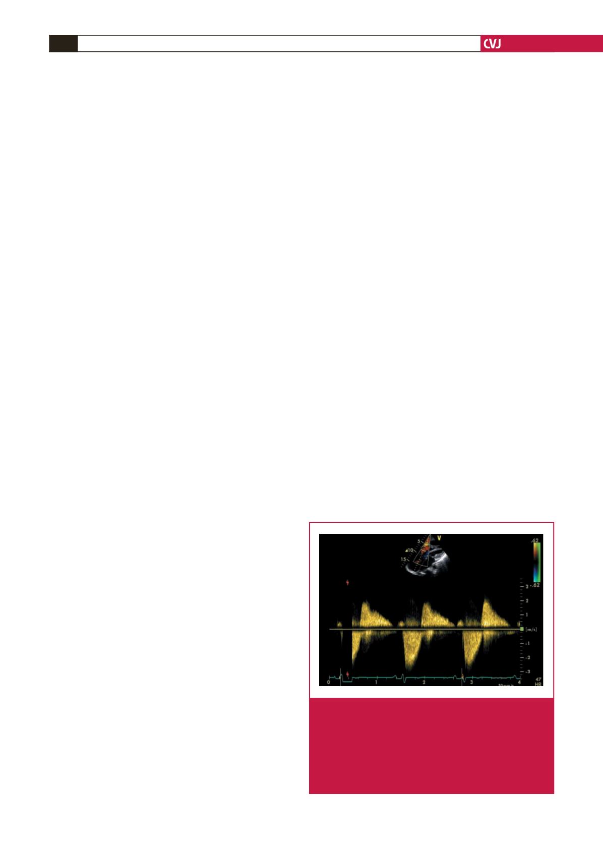
Fig. 4.
Continuous-wave Doppler demonstrating a dagger-
shaped pattern in a patient with severe tricuspid regur-
gitation due to carcinoid heart disease, with early peak
pressure and a rapid decline. In addition, the patient
had marked failure of coaptation (FOC) resulting
from severely thickened and retracted tricuspid valve
leaflets due to underlying carcinoid disease (image
courtesy of Fox and Khattar).
5