


 13 / 78

CARDIOVASCULAR JOURNAL OF AFRICA • Volume 30, No 1, January/February 2019
AFRICA
11
of PH using an estimated PASP cut-off value of
>
36 mmHg was
present in 85 (38.8%) subjects.
Clinical characteristics of HF subjects with and without PH are
summarised in Table 2. HF subjects with PH tended to be male (
p
=
0.03) with a lower body mass index (BMI) (
p
=
0.002) and higher
NYHA functional class (
p
<
0.001) compared with those without
PH. However, there was no significant difference in the frequency
of previous hospitalisations (
p
=
0.74) and duration of HF (
p
=
0.26) between the groups. The presence of PH was significantly
associated with a HF aetiology of dilated cardiomyopathy (
p
=
0.02) and valvular heart disease (
p
=
0.015), while the absence of PH
was associated with hypertensive heart disease (
p
=
0.008) (Table 3).
Comparison of the echocardiographic characteristics of HF
subjects with and without PH is summarised in Table 4. HF
subjects with PH had significantly higher left atrial and LV
chamber dimensions (
p
<
0.001). They also had poorer LV
systolic function, higher LV diastolic dysfunction grade (Fig. 1)
and LV filling pressures, assessed by E/e
′
ratio (
p
<
0.001 for all).
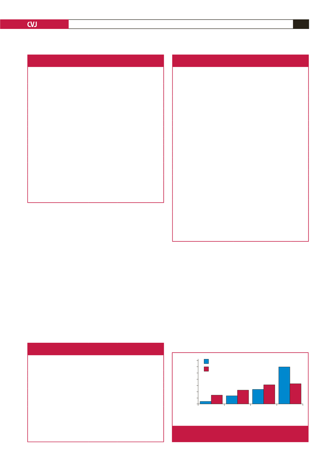
The presence of PH was associated with more severe MR (Fig. 2).
The subjects with PH had significantly worse parameters of right
heart structure and function, compared with the non-PH group.
The age and BMI of the study population correlated negatively
with PASP (
p
=
0.02,
p
<
0.001, respectively). The pulse of the
subjects had a significant but weak positive correlation with
PASP (
r
=
0.138,
p
=
0.04). Other clinical parameters did not
show significant correlations with PASP (Table 5).
PASP correlated significantly with all selected echocardio-
graphic parameters of left heart structure and function, such
as left atrial volume index (LAVI), LV mass index, LVEF and
E/e
′
ratio (Table 5). The mitral regurgitant volume correlated
positively with PASP (
r
=
0.269,
p
<
0.001). All parameters of RV
structure and function correlated significantly with PASP.
Table 2. Comparison of the clinical characteristics of subjects
with and without pulmonary hypertension
Parameters
PH group
(
n
=
85)
Non-PH group
(
n
=
134)
p-
value
Gender (M/F, %)
69/31
55/45
0.028
Age (years)
55.0
±
13.9
57.2
±
14.9 0.28
BMI (kg/m
2
)
25.8
±
5.1
28.1
±
5.7
0.002
Pulse (beats/min)
85.2
±
14.6
80.9
±
13.7 0.03
SBP (mmHg)
114
±
19.9
119
±
20.3 0.05
DBP (mmHg)
75.7
±
14.0
74.5
±
12.8 0.52
NYHA class,
n
(%)
<
0.001
II
29 (34.1)
102 (76.1)
III
41 (48.2)
27 (20.1)
IV
15 (17.6)
5 (3.7)
Previous admissions,
n
(%)
0.74
None
38 (44.7)
65 (48.5)
1
31 (36.5)
42 (31.3)
≥ 2
16 (18.8)
27 (20.1)
Duration of heart failure (weeks)*
0.26
1–25
25 (32.9)
38 (33.3)
26–104
31 (40.8)
35 (30.7)
105–579
20 (26.3)
41 (36.0)
*Grouped as tertiles; PH, pulmonary hypertension; BMI, body mass index;
SBP, systolic blood pressure; DBP, diastolic blood pressure; NYHA, New York
Heart Association.
Table 3. Comparison of heart failure aetiologies
among subjects with and without PH
Parameters
PH group
(
n
=
85)
n (%)
Non-PH group
(
n
=
134)
n (%)
p-
value
Hypertensive heart disease
32 (37.6)
75 (56.0)
0.008
Idiopathic dilated cardiomyopathy 37 (43.5)
37 (27.6)
0.02
Peripartum cardiomyopathy
3 (3.5)
11 (8.2)
0.26
Valvular heart disease
8 (9.4)
2 (1.5)
0.02
RCM/amyloid heart disease*
4 (4.7)
1 (0.7)
0.08
Ischaemic heart disease
1 (1.2)
3 (2.2)
0.96
Obesity-related heart disease**
0 (0)
4 (3.0)
0.16
PH, pulmonary hypertension, RCM, restrictive cardiomyopathy.
*This was diagnosed by suggestive echo findings of severe LV wall thickening,
myocardial speckled appearance and normal or low-voltage limb lead voltages
on 12-lead ECG.
** This was a diagnosis of exclusion made in subjects with a BMI
>
30 kg/m
2
and no other other identifiable cause or explanation of heart failure.
Table 4. Comparison of echocardiographic characteristics of subjects
with and without pulmonary hypertension
Parameters
PH group
(
n
=
85)
Non-PH group
(
n
=
134)
p-
value
LV diastolic diameter (cm)
6.3
±
1.1
5.7
±
1.3
<
0.001
LV systolic diameter (cm)
5.2
±
1.2
4.4
±
1.4
<
0.001
LA volume index (ml/m
2
)
89.8
±
72.9
54.7
±
25.9
<
0.001
Fractional shortening (%)
17.3
±
8.4
22.6
±
10.0
<
0.001
Ejection fraction (%)
35.4
±
15.0
44.1
±
16.7
<
0.001
LV mass index (g/m
2
)
172
±
54.9 138.5
±
53.4
<
0.001
TAPSE (cm)
1.6
±
0.4
2.0
±
0.5
<
0.001
RV basal diameter (cm)
5.0
±
0.8
4.2
±
0.8
<
0.001
RV wall thickness (cm)
0.37
±
0.16
0.33
±
0.09
<
0.04
RA area (cm
2
)
28.8
±
7.7
19.4
±
6.2
<
0.001
Eccentricity index*
1.12
±
0.18
1.03
±
0.11
<
0.001
Presence of D-sign,
n
(%)
26 (30.6)
7 (5.2)
<
0.001
E/e
′
ratio
15.9
±
5.4
11.7
±
5.5
<
0.001
Diastolic dysfunction grade
<
0.001
1 or normal
3 (3.6)
55 (41.0)
2
13 (15.7)
36 (26.9)
3
67 (80.7)
5 (32.7)
MR severity,
n
(%)
<
0.001
mild
11 (12.9)
30 (22.4)
moderate
20 (23.5)
41 (30.6)
severe
51 (60.0)
44 (32.8)
Mitral regurgitant volume (ml)
85.8
±
64.7
67.4
±
59.3
<
0.04
PH, pulmonary hypertension; LA, left atrium; LV, left ventricle; TAPSE, tricus-
pid annular plane systolic excursion; RVWT, right ventricular wall thickness; E
velocity, mitral inflow E velocity; e
′
, early mitral annular diastolic velocity; MR,
mitral regurgitation.
*Eccentricity index is the ratio of the LV anteroposterior and septolateral diam-
eters measured in the parasternal short-axis view.
Mild MR (MRvol < 30 ml), moderate (MRvol 30–59 ml),
severe MR (MRvol > 60 ml)
No MR Mild MR Moderate MR Severe MR
Percent
70
60
50
40
30
20
10
0
PH group (PASP > 36 mmHg)
non-PH group
Fig. 1.
Severity of mitral regurgitation in the PH and non-PH
subgroups.