CARDIOVASCULAR JOURNAL OF AFRICA • Vol 23, No 3, April 2012
AFRICA
e1
Case Report
Iatrogenic hydropneumopericardium
F PETERS, A PATEL, R ESSOP
Abstract
We report a case of iatrogenic hydropneumopericardium
in a patient with diffuse scleroderma. The transthoracic
echocardiogram revealed classical features of this condition.
This case resolved spontaneously after five days using only
60
%
oxygen, which may have augmented resolution in this
instance.
Keywords:
iatrogenic, hydropneumopericardium, scleroderma
Submitted 7/7/10, accepted 15/4/11
Cardiovasc J Afr
2012;
23
: e1–e2
DOI: 10.5830/CVJA-2011-014
Case report
A 47-year-old woman with diffuse scleroderma was found
on chest X-ray to have cardiomegaly and was referred for an
echocardiogram. The examination of the cardiovascular system
was normal except for the presence of severe uncontrolled
hypertension. Her blood results were normal, including her renal
function.
The transthoracic echocardiogram revealed normal
left ventricular systolic function, concentric left ventricular
hypertrophy with a large pericardial effusion, accompanied by
diastolic collapse of the right ventricular free wall and systolic
collapse of the right atrium. The rest of the heart and aorta were
normal.
Echo-guided pericardiocentesis using the subxiphoid
approach was done to drain 800 ml of serous fluid. This exudate
had normal adenosine deaminase levels, as well as cytology and
microbiological analysis.
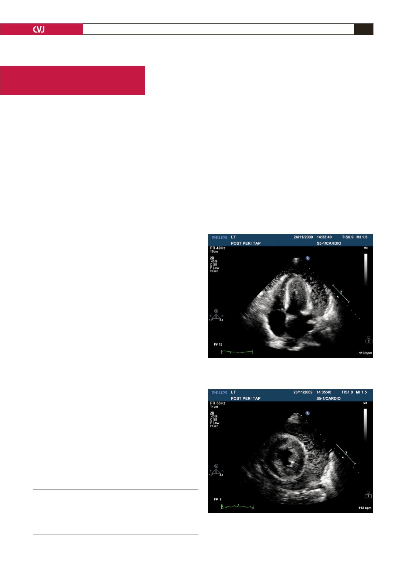
A transthoracic echocardiogram performed the next day
revealed a large pericardial effusion with multiple swirling
‘hyperdensities’within the fluid with no echo signs of tamponade
(Figs 1, 2). A chest X-ray confirmed a large pneumopericardium
(Fig. 3).
A CT scan of the thorax and upper abdomen with hexabrix
follow-through confirmed the hydropneumopericardium and
revealed no communication with the oesophagus, lungs and
stomach. The mediastinum was normal and there was no air in
the subcutaneous tissue of the chest.
The patient remained stable in the intensive care unit and
was put on a 60
%
oxygen face mask. The tract where the
pericardiocentesis was performed appeared to have closed
spontaneously. An echocardiogram four days later showed almost
complete resolution of the air bubbles, with a large residual
pericardial effusion (Fig. 4). The patient was subsequently
started on steroids and remained stable. She was followed up as
an outpatient, with partial resolution of her pericardial effusion
on discharge.
After investigation we concluded that this patient’s pericardial
Division of Cardiology, Chris Hani Baragwanath Hospital,
Johannesburg, South Africa
F PETERS, MB BCh, FCP (SA), Cert Cardiology (SA), ferande.
A PATEL, MB BCh, FCP (SA)
R ESSOP, MB BCh, FCP (SA), FACC, FRCP (London)
Fig. 1. Apical four-chamber view demonstrating the
swirling echogenic bubbles within the large pericardial
effusion.
Fig. 2. Parasternal short-axis view demonstrating the
large pericardial effusion within which the swirling echo-
genic bubbles can be seen.


