CARDIOVASCULAR JOURNAL OF AFRICA • Vol 23, No 3, April 2012
AFRICA
e9
Very late thrombosis of a paclitaxel-eluting stent after
72 months in a patient on dual anti-platelet therapy
V SUBBAN, L KALIDOSS, MA SANKARDAS
Abstract
Very late thrombosis continues to be a major cause of
concern in the era of drug-eluting stents. The duration of
vulnerability to this complication remains undefined. A
62-year-old diabetic male underwent primary percutaneous
coronary intervention with a Taxus Express stent (Boston
Scientific, Natick, Mass) implantation in 2003 for anterior
wall myocardial infarction (AWMI). The patient was on dual
anti-platelet treatment. He was asymptomatic and his stress
test was negative in 2008. After 72 months, the patient was
admitted with acute AWMI resulting from stent thrombosis,
which was treated successfully. This case underscores the
importance of realising that very late stent thrombosis may
occur when patients present with angina symptoms.
Keywords:
very late stent thrombosis, drug-eluting stent, Taxus
Express stent
Submitted 29/12/10, accepted 31/5/11
Cardiovasc J Afr
2012;
23
: e9–e11
DOI: 10.5830/CVJA-2011-022
Drug-eluting stents (DES) are widely used in contemporary
practice and this has made percutaneous coronary intervention
(PCI) an accepted treatment for diabetic patients and those
with complex coronary artery disease.
1
Drug-eluting stents
effectively suppress neo-intimal hyperplasia and resulting
restenosis, but inadequate stent endothelial coverage results in
late stent thrombosis (LST). The exact period of time required
for complete neo-intimal healing and susceptibility to stent
thrombosis is largely unknown.
2
Here we report a case of very
late stent thrombosis 72 months after primary percutaneous
intervention (primary PCI) to the left anterior descending
coronary artery (LAD).
Case report
A 62-year-old diabetic male suffered an anterior wall ST-elevation
myocardial infarction in 2003. The patient was taken up for
primary PCI. The angiogram showed the right coronary artery
with no significant stenosis and a totally occluded LAD after the
first septal branch.
The LAD lesion was pre-dilated with a 2
×
10-mm Maverick
balloon (Boston Scientific Corporation, Natick, Massachusetts)
and then a 2.75
×
32-mm Taxus Express stent (Boston Scientific
Corporation, Natick, Massachusetts) was implanted, with good
angiographic results.
The peri-procedural period was uneventful. The patient was
continued on aspirin, clopidogrel and statin. The ejection fraction
with echocardiography was 48% on discharge. The patient
was on regular follow up with an indefinite dual anti-platelet
regimen. His stress test was negative for inducible ischaemia in
2008.
The patient presented to the emergency department in
October 2009 with a history of chest discomfort of two hours’
duration. The electrocardiogram showed ST-segment elevation
fromV1
-
V6. The echocardiogram revealed regional wall motion
Institute of Cardio-Vascular Diseases, Madras Medical
Mission, Chennai, India
V SUBBAN, MD, DM,
L KALIDOSS, MD, DM
MA SANKARDAS, MD, DM
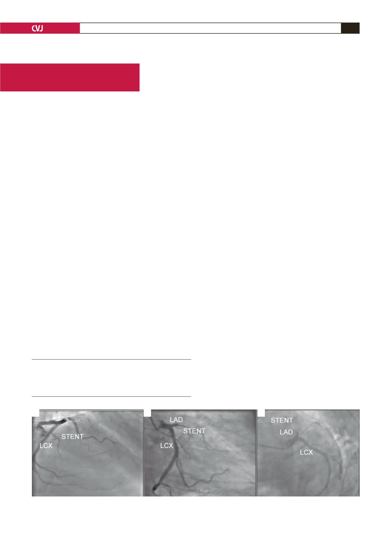
A
B
C
Fig. 1. Left coronary artery angiogram in the antero-posterior cranial (A), right anterior oblique caudal (B), and left
anterior oblique caudal (C) views, showing the totally occluded left anterior descending coronary artery at the proxi-
mal end of the stent.
Case Report


