CARDIOVASCULAR JOURNAL OF AFRICA • Vol 23, No 3, April 2012
e12
AFRICA
Post-infarction myocardial rupture: a case of pericardial
tamponade salvaged by auto-blood transfusion
AK KHALEDI, F LARTI, S SAFARI
Left ventricular free-wall rupture (LVFWR) is a serious and
lethal complication of acute myocardial infarction. Although
this complication is not common, the fatality rate is high due
to haemodynamic collapse in the setting of cardiac tampon-
ade. We report a case of LVFWR in a patient with a rare
blood group, who survived because of an innovative tech-
nique for pericardiocentesis and simultaneous transfusion of
the aspirated blood into the femoral sheath. A video of the
patient’s ventriculography is provided.
Keywords:
myocardial rupture, auto-transfusion, cardiac
tamponade, myocardial infarction
Submitted 3/5/10, accepted 1/6/11
Cardiovasc J Afr
2012;
23
: e12–e13
DOI: 10.5830/CVJA-2011-026
Case report
A 65-year-old man presented to the emergency department
(ED) with a history of retrosternal chest pain of some hours
before admission. His condition was stable. The ECG showed
non-significant ST-T changes. With a working diagnosis of acute
coronary syndrome, the patient was transferred to the critical
care unit. His cardiac enzymes were normal. The standard
medical therapy for unstable angina was started.
The patient’s ECG on the second day of admission showed
evidence of evolving inferior myocardial infarction (MI). An
echocardiography showed an ejection fraction of 50% and mild
infrobasal hypokinesia. On the third day, an exercise tolerance
test (ETT) was negative until the end of stage III of the Bruce
protocol. The patient was discharged one day after the normal
ETT with a recommendation of undergoing elective angiography
if he desired.
He came back to the ED one day after discharge, with
typical chest pain. His condition was stable. This time the ECG
showed reappearance of ST elevation in the inferior leads. The
patient received streptokinase (SK) and his chest pain resolved.
Nine hours later, the patient became agitated and hypotensive
(BP
=
70/50 mmHg). Immediately bedside echocardiography
was performed and massive pericardial effusion with cardiac
tamponade was detected.
Cardiac surgeonswereconsultedand thepatientwas transferred
to the catheterisation laboratory for pericardiocentesis. Because
of the patient’s rare blood group, the blood bank could not afford
even one unit of packed cells and required at least one hour for
blood preparation. An arterial femoral sheath was inserted, both
for performing coronary angiography and auto-transfusion of the
blood aspirated from the pericardial sac.
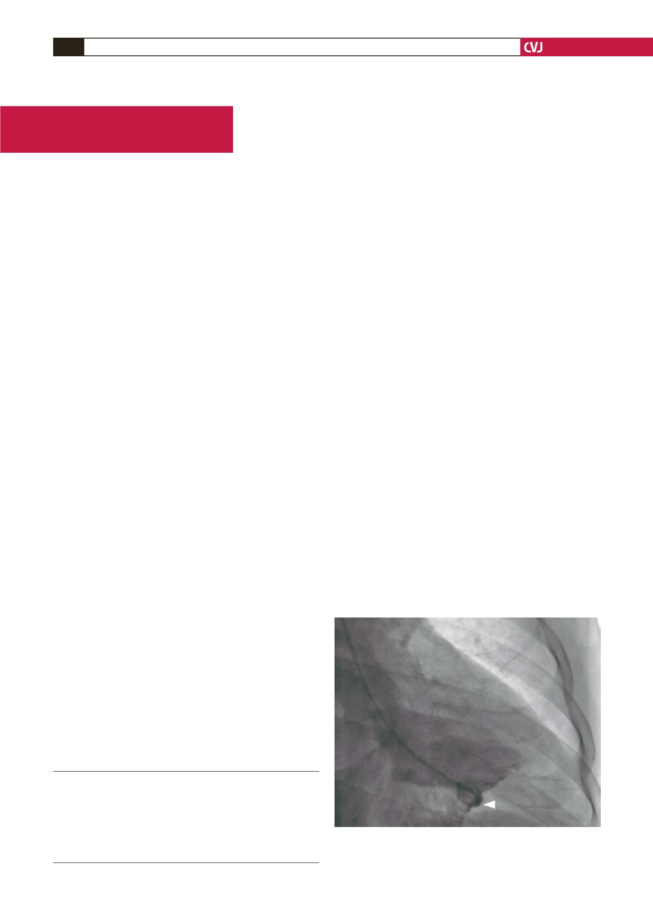
The patient’s ventriculography is shown in Fig. 1 and
a supplementary video is provided at
journal/vol23/vol23_issue3/videos/DOI-10-5830-CVJA
-2011-026.php. Coronary angiography showed triple-vessel
disease: the left anterior descending artery (LAD) had 50%
stenosis after the first diagonal branch, the left circumflex
artery (LCX) had 80% stenosis after the second obtuse marginal
branch, and the right coronary artery (RCA) was totally cut off
after the acute marginal branch.
The patient underwent emergency coronary artery bypass
graft (CABG) surgery using cardiopulmonary bypass, and the
ventricular free-wall rupture was repaired with a synthetic patch.
After three days in the intensive care unit and a total hospital
stay of two weeks, the patient was discharged home in a good
condition. At the 18-month follow-up visit, he was still doing
well.
Department of Cardiology, Imam Khomeini Hospital, Tehran
University of Medical Sciences, Tehran, Iran
AK KHALEDI, MD
F LARTI, MD,
Department of General Surgery, Imam Khomeini Hospital,
Tehran University of Medical Sciences, Tehran, Iran
S SAFARI
Fig 1. Left ventriculogram showing entrance of contrast
media into the pericardial sac. This indicates a free-wall
rupture (arrowhead).
Case Report


