CARDIOVASCULAR JOURNAL OF AFRICA • Vol 23, No 3, April 2012
AFRICA
e7
(Diltiazem) and aspirin. He has remained well and asymptomatic
for 22 months.
Discussion
In adolescents, the pathogenesis of STEMI is unclear and often
different from that of adult STEMI.
1
The coronary arteries are
usually normal in young patients. The pathogenetic mechanisms
may be coronary vasospasm, hypercoagulable state secondary
to hereditary thrombophilia, collagen vascular disease and
embolisation. However, in most reported cases there is no
obvious cause.
1-3
Prothrombotic factors may also contribute to the development
of STEMI at a young age.
1-3
In our case, we observed multiple
gene polymorphisms, such as heterozygote beta fibrinogen
(455G
>
A), MTHFR (C677T and A1298C) and CETP (TaqI
β
),
which most likely predisposed to the prothrombotic state. In
addition, PAI-1 4G/4G and HPA1 a/a genotypes were also
present.
It has been reported that heterozygous mutation in the beta
fibrinogen gene can be associated with STEMI and coronary
artery disease.
5,6
Similarly, the reduced activity of MTHFR
may predispose to coronary events.
4,7-9
However, it is unclear if
there is a significant association between two common MTHFR
mutations (C677T and A1298C) and hyperhomocysteinaemia.
7
The homocysteine level was normal in our case. On the
other hand, C677T and A1298C MTHFR mutations have been
reported to be associated with acute STEMI in adolescents and
young adults.
4,7-9
CETP mediates the transfer of cholesteryl esters and
triglycerides among lipoprotein particles and plays a crucial
role in reverse cholesterol transport. The Taq1
β
polymorphism
of CETP may be associated with the early onset of STEMI and
coronary atherosclerosis, independent of high-density lipoprotein
cholesterol levels.
10
The PAI-1 4G/4G genotype may increase PAI-1 levels and/or
activity, especially in young males.
3,9,11
Over-expression of PAI-1
may induce thrombosis and vulnerability to atherosclerotic
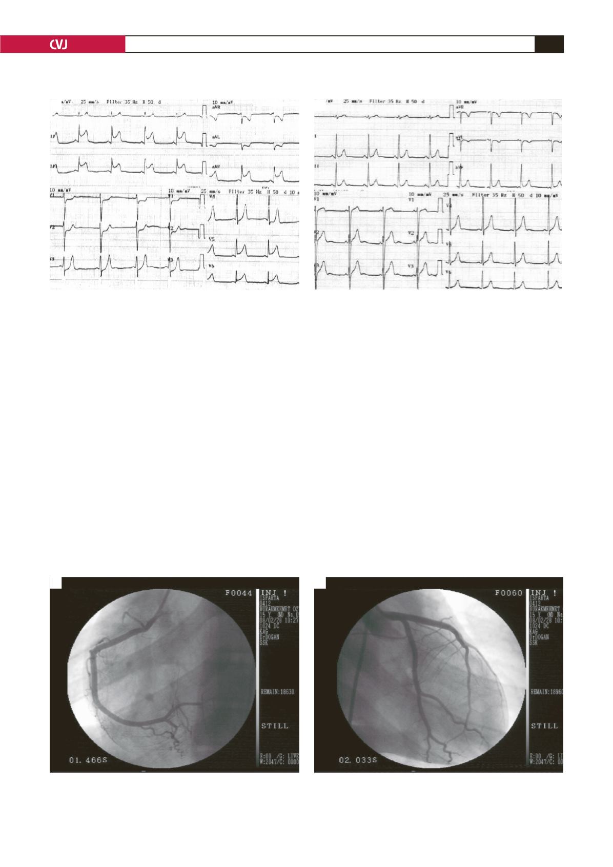
Fig. 1. A: Electrocardiogram shows ST-segment elevation in leads II, III-aVF and V5-6, and ST-segment depression in
leads aVL and V1-3 . B: At follow up, these changes had disappeared.
B
A
Fig. 2. A: Coronary angiogram demonstrates the right coronary artery with no stenosis after intracoronary nitroglycer-
in injection. B: The left coronary system appears to be normal. However, there was stasis and slow-flow phenomenon
in the proximal segment of the left anterior descending artery.
B
A


