


 15 / 67

CARDIOVASCULAR JOURNAL OF AFRICA • Vol 24, No 2, March 2013
AFRICA
13
demonstrated by the significantly smaller infarct size observed
(Fig. 3). The effect of this treatment was highly significant (
p
<
0.001,
n
=
9 per group) as indicated by two-way ANOVA.
Analyses of proteins forming part of the insulin-
signalling cascade
Protection against myocardial damage induced by ischaemia–
reperfusion and culminating in the formation of an infarct has
been ascribed, among others, to the activity of the phosphatidyl-
inositol-3-kinase (PI-3K) pathway. In view of the previously
reported improvements in insulin sensitivity of cardiomyocytes,
induced by
P
glandulosa
treatment,
8
we systematically analysed
the proteins involved in this signalling cascade.
As summarised in Table 2 and shown in Fig. 4, hearts
from the DIO animals presented with a significantly lower
phosphorylated:total ratio of the central protein in this cascade,
protein kinase B or Akt. This ratio was significantly improved
by treatment. In addition, the expression of the p85 regulatory
subunit of the PI-3K enzyme was significantly lower in hearts
from the DIO animals, whereas this was not the case after
treatment.
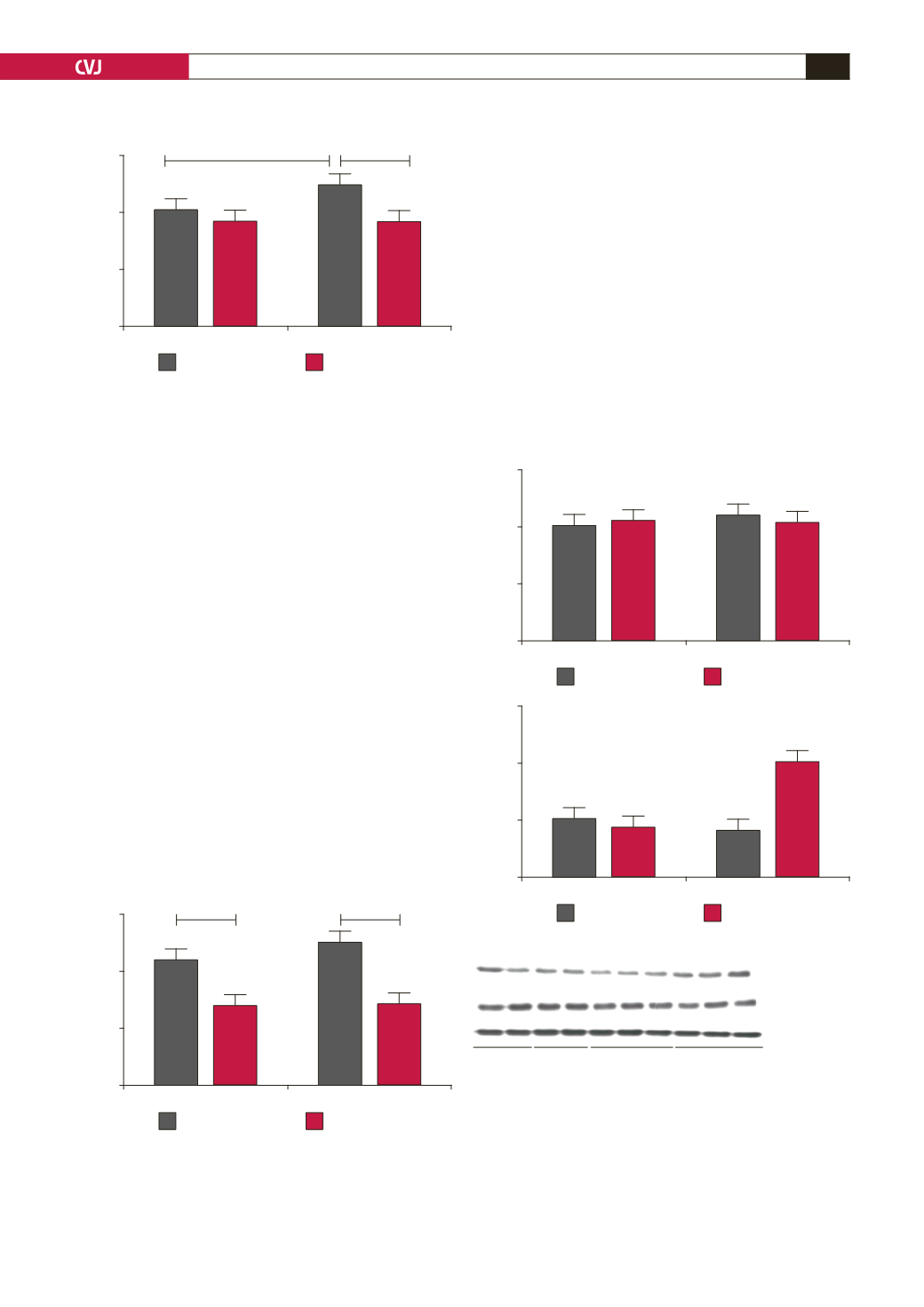
Fig. 2. After the 16-week diet plus
P glandulosa
treatment,
isolated hearts from DIO rats were perfused
ex vivo
in
the working-heart mode. They were subjected to regional
ischaemia as described in Methods. Infarct size was
determined as a percentage of the area at risk of infarc-
tion. *
p
<
0.05, **
p
<
0.01,
n
=
15–17 per group.
60
40
20
0
Control
DIO
Infarct size (% of area at risk)
minus treatment
plus treatment
*
**
Fig. 3. After the eight weeks of treatment, hearts were
removed from the CIRKO mice and perfused
ex vivo
in the
Langendorff mode and subjected to NICA as described in
Methods. Infarct size was determined throughout the
whole heart and expressed as a percentage of the total
surface. **
p
<
0.01, ***
p
<
0.001,
n
=
9 per group.
60
40
20
0
Control
CIRKO
Infarct size
minus treatment
plus treatment
***
**
Treatment also resulted in a lower expression of the
phosphatase and tensin homologue deleted on chromosome 10
(PTEN) with a higher state of phosphorylation of this enzyme
(Fig. 5). Phosphorylation of PTEN further inactivates this
enzyme, responsible also for the dephosphorylation of PKB/
Akt.
17,18
Anti-hypertensive effects
As the DIO diet does not cause high blood pressure, we used
a modification of a high-fat diet to induce hypertension in the
animals.
12
As can be seen in Fig. 5, these animals developed a
significant elevation of their blood pressure within four weeks
(HFD 135.88
±
2.0 vs control 125.85
±
1.9 mmHg,
p
<
0.05,
n
=
8 per group).
Fig. 4. Hearts from the treated and untreated DIO
animals were removed without any intervention and
stored in liquid nitrogen. Tissue lysates were prepared
and Western blotting was performed as described in
Methods. A: bar charts of the expression of PKB protein
as well as the ratio of phosphorylated vs total protein.
*
p
<
0.05 vs control; **
p
<
0.01 vs untreated DIO,
n
=
6
individual hearts analysed per group. B is a representa-
tive blot depicting these proteins and beta-tubulin, used
as an indicator of equal loading.
1.5
1.0
0.5
0.0
3
2
1
0
Control
DIO
Control
Diet
Arbitrary densitrometry units
Ratio (phospho/total)
minus treatment
minus treatment
plus treatment
plus treatment
A
B
*
**
Beta-tubulin
Phospho-PKB
Total PKB
Contr Contr + Tr
DIO + Tr
DIO