


 64 / 67

CARDIOVASCULAR JOURNAL OF AFRICA • Volume 26, No 2, March/April 2015
e10
AFRICA
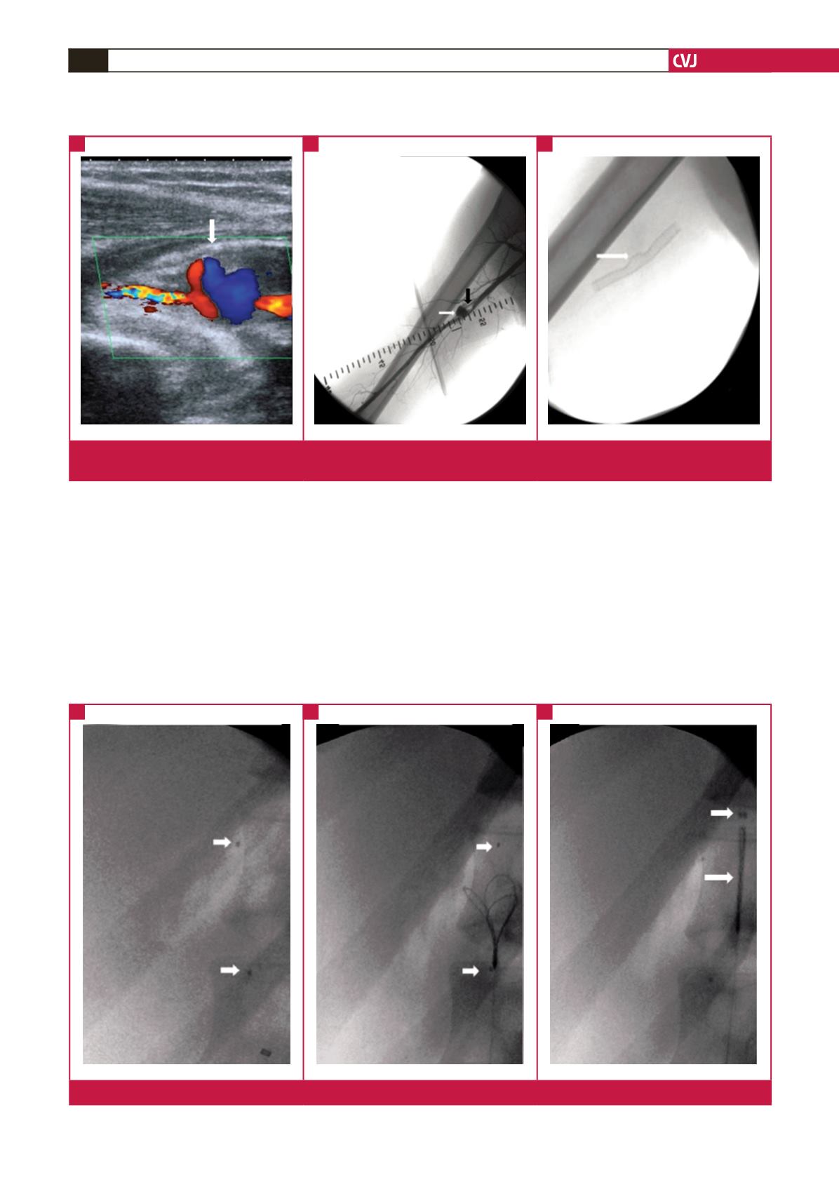
checked the instruments and observed that the distal part of the
balloon catheter was broken. Angiography showed the catheter
tip in the abdominal aorta (Fig. 2A). In the same session,
the catheter piece was successfully removed using a three-
dimensional snare device (Mini EN snare, Gainsville, FL, USA).
Control angiography showed the piece was removed completely
(Figs 2B, C and 3).
Discussion
Pseudo-aneurysms may develop when the layers of an artery are
disrupted and a haematoma forms within the peri-arterial tissues.
The haematoma becomes a cavity of blood and thrombus, and
this becomes a pseudo-aneurysm if it communicates with the
true vessel lumen. There are many aetiologies but pseudo-
aneurysms are primarily caused by anastomosis dehiscence,
vascular procedures, and blunt or penetrating trauma.
1
Endovascular graft implantation is a new, minimally
invasive intervention. It can be used for aneurysms and pseudo-
aneurysms of the peripheral arterial system and for arteriovenous
fistulae. With the introduction of endovascular techniques for
applications related to vascular injury, less-invasive modalities
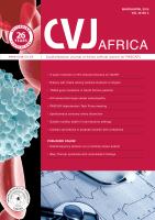
Fig. 1.
A: Colour Doppler USG showing brachial artery pseudo-aneurysm sac, B: white arrow is DSA angiographically visualised
brachial artery pseudo-aneurysm sac, black arrow is haematoma at the proximal segment of the sac, C: implanted stent.
A
B
C
Fig. 2.
A: Removal of broken catheter piece, B, C: three-dimensional snare device.
A
B
C