


 59 / 64

CARDIOVASCULAR JOURNAL OF AFRICA • Volume 27, No 1, January/February 2016
AFRICA
e1
Case Report
Localised pericardial effusion mimicking anterior
myocardial infarction following coronary angiography
Aynur Acibuca, Demet Menekse Gerede, Veysel Ozgur Baris, Mustafa Kilickap
Abstract
The diagnosis of pericarditis is important, especially in patients
assumed to have acute coronary syndrome. Distinguishing
these two conditions is vital but not always easy. Accurate
diagnosis is essential to provide appropriate treatment as soon
as possible and to avoid inappropriate invasive procedures. By
highlighting this distinction, we report a case of pericarditis
that occurred after percutaneous coronary intervention and
mimicked acute coronary syndrome.
Keywords:
regional pericarditis, myocardial infarction, acute
stent thrombosis, located pericardial effusion
Submitted 3/8/15, accepted 14/11/15
Cardiovasc J Afr
2016;
27
: e1–e3
www.cvja.co.zaDOI: 10.5830/CVJA-2015-086
Regional pericarditis has been described but remains a
relatively unknown and under-diagnosed condition. There are
no electrocardiography (ECG) criteria to diagnose regional
pericarditis and only a few studies have investigated regional
pericarditis. Although regional pericarditis is usually observed
in patients following myocardial infarction (MI), it has also been
reported in other conditions.
1,2
We present a case of regional
pericarditis with electrocardiographic features mimicking
anterior MI.
Case report
A 58-year-old male smoker presented with a one-month history
of exercise chest pain. His exercise ECG was borderline normal,
so coronary angiography (CAG) was performed. The CAG
revealed severe stenosis in the circumflex and right coronary
artery (RCA). Borderline severe stenosis was also detected in the
left anterior descending (LAD) coronary artery (Figs 1, 2). The
fractional flow reserve value was 0.92.
Department of Cardiology, Ankara University School of
Medicine, Ankara, Turkey
Aynur Acibuca, MD,
aynuracibuca85@gmail.comDemet Menekse Gerede, MD
Veysel Ozgur Baris, MD
Mustafa Kilickap, MD
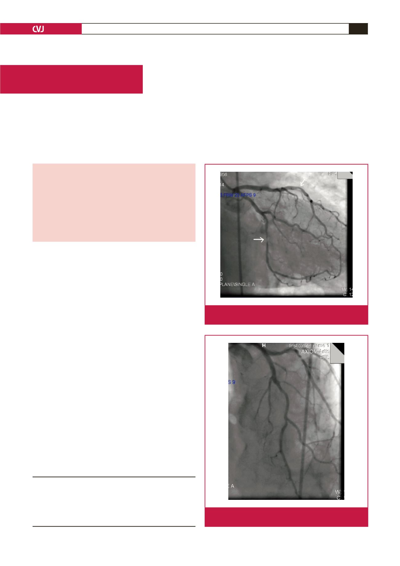
Fig. 1.
The right anterior oblique view shows stenosis in the
circumflex and left anterior descending coronary artery.
Fig. 2.
The anteroposterior view shows stenosis in the circum-
flex and left anterior descending coronary artery.