CARDIOVASCULAR JOURNAL OF AFRICA • Vol 23, No 6, July 2012
e12
AFRICA
Case Report
Dilated cardiomyopathy secondary to coarctation of the
aorta was completely resolved after stent implantation
MUSTAFA TARIK AĞAÇ, ZEYDIN ACAR, RAMAZAN AKDEMIR, LEVENT KORKMAZ, ABDÜLKADIR KIRIŞ,
ALI RIZA AKYÜZ, HAKAN ERKAN
Abstract
Although rare in adults, coarctation of the aorta can present
as an occult cause of cardiomyopathy. In this case report, we
describe a 55-year-old man who presented to our clinic with
new-onset symptoms of heart failure. Transthoracic echo-
cardiography showed a dilated left ventricle with severely
depressed systolic function. Cardiac catheterisation showed
no critical coronary artery disease but a severe aortic coarc-
tation just distal to the origin of the left subclavian artery,
with a peak-to-peak systolic pressure gradient of 40 mmHg.
We successfully implanted a balloon-expandable stent with
good angiographic results. The procedure resulted in imme-
diate clinical improvement. Six-months after stent implanta-
tion, the patient was free of symptoms and an echocardio-
graphic examination showed completely normalised cardiac
size and systolic function.
Keywords:
aortic coarctation, dilated cardiomyopathy, heart
failure, stent implantation
Submitted 12/2/11, accepted 21/9/11
Cardiovasc J Afr
2012;
23
: e12–e13
DOI: 10.5830/CVJA-2011-061
In patients referred for evaluation of heart failure, it is
important to exclude reversible causes. This includes
consideration of mechanical causes, such as aortic stenosis,
which may produce a clinical picture resembling dilated
cardiomyopathy, for which surgical correction is possible.
Similarly, increased afterload in the setting of aortic
coarctation may produce heart failure.
1,2
In the present report,
we describe an adult patient presenting in the fifth decade
of life with dilated cardiomyopathy secondary to occult,
congenital coarctation of the aorta, who recovered completely
after stent implantation.
Ahi Evren Chest, Heart and Vascular Surgery Hospital,
Soğuksu Mah, Trabzon, Turkey
MUSTAFA TARIK AĞAÇ, MD,
ZEYDIN ACAR, MD
RAMAZAN AKDEMIR, MD
LEVENT KORKMAZ, MD
ABDÜLKADIR KIRIŞ, MD
ALI RIZA AKYÜZ, MD
HAKAN ERKAN, MD
Case report
A 55-year-old man was admitted to our hospital with a
one-week history of increasing dyspnoea, orthopnoea and
nocturnal dyspnoea. He had noticed a gradual limitation in
exercise tolerance over the previous two years. His medical
history revealed only systemic arterial hypertension of 22 years’
duration, which was poorly controlled despite concurrent use of
three antihypertensive medications.
On physical examination, his blood pressure was 190/100
mmHg. There was a differential blood pressure of 30 mmHg
between the upper and lower extremities. There were basal rales
in the lower lung fields and auscultation showed rapid heart
sounds with S3 gallop. A harsh II/VI systolic murmur was heard
along the left scapular border.
Echocardiography revealed a dilated left ventricle with
end-diastolic (LVEDD) and end-systolic diameters (LVESD)
of 60 and 53 mm, respectively. Left ventricular contraction
was diffusely hypokinetic, with a calculated ejection fraction
(EF) of 30% (Video 1). Continuous-wave Doppler interrogation
revealed a peak velocity of 2.9 m/s along the descending
aorta. No significant valvular dysfunction was noted. Cardiac
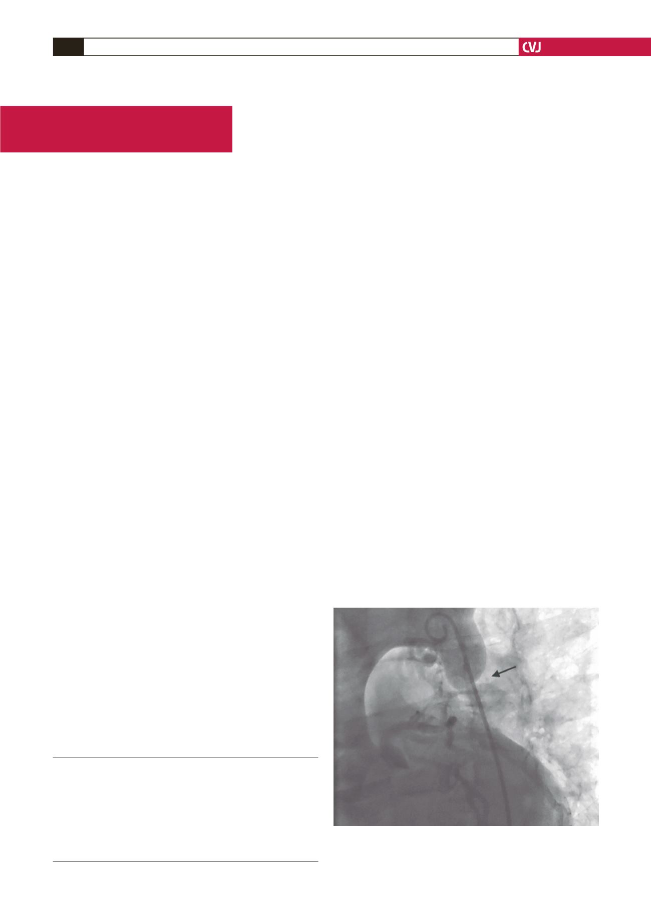
catheterisation showed normal coronary arteries, but a severe
funnel-shaped aortic coarctation was detected just distal to the
origin of the left subclavian artery (Fig. 1, Video 2) with a peak-
to-peak systolic pressure gradient of 40 mmHg.
Fig. 1. Aortography in the left anterior oblique view
showing severe funnel-shaped aortic coarctation just
distal to the origin of the left subclavian artery.


