CARDIOVASCULAR JOURNAL OF AFRICA • Vol 23, No 6, July 2012
e14
AFRICA
Case Report
A rare case of spontaneous rectus sheath haematoma
in a patient with mechanical prosthetic aortic and mitral
valves
AHMET AYKAN, ALİ OGUZ, MUSTAFA YİLDİZ, MEHMET ÖZKAN
Abstract
Every year nearly 300 000 patients have heart valve opera-
tions and mostly prosthetic valves are inserted. Coumadin
is the mainstay of therapy in these individuals but it has
many side effects, mostly related to its anticoagulant effect.
Rectus sheath haematoma (RSH) is a rare complication of
abdominal trauma, surgery and excessive strain, however,
anticoagulant agents may predispose to this condition with-
out any precipitating event. Reversal of anticoagulation and
resuscitation with fluids and blood products are necessary
but anticoagulation is crucial in patients with prosthetic
valves, as they have acquired thrombotic diathesis. Herein
we report on a case of spontaneous RSH in a patient with
prosthetic mitral and aortic valves and a history of prosthetic
valve thrombosis. He was successfully managed medically.
Keywords:
rectus sheath, haematoma, prosthetic valve, warfarin
Submitted 23/3/11, accepted 22/11/11
Cardiovasc J Afr
2012;
23
: e14–e15
DOI: 10.5830/CVJA-2011-070
Rectus sheath haematoma (RSH), secondary to abdominal
trauma, surgery and excessive strain, is a rare complication.
Its incidence without any precipitating event is increasing
with the growing use of antiplatelet and anticoagulant agents.
1
Management of RSH in patients with prosthetic mechanical
heart valves is a challenge as anticoagulation in these subjects
is crucial, whereas keeping them on anticoagulation may cause
death.
Case report
A 36-year-old male patient was admitted to the emergency
department with a two-day history of abdominal pain, poor
appetite, dizziness, fatigue and discolouration of the abdomen
and flank. He had had no recent trauma or surgery. His bowel
habits were normal and there was no discolouration of the faeces
or urine.
He had had mechanical prosthetic mitral and aortic valve
surgery for rheumatic heart disease five years earlier and had
a history of prosthetic mitral valve thrombosis, which was
successfully treated with thrombolytic therapy. He was on
coumadin.
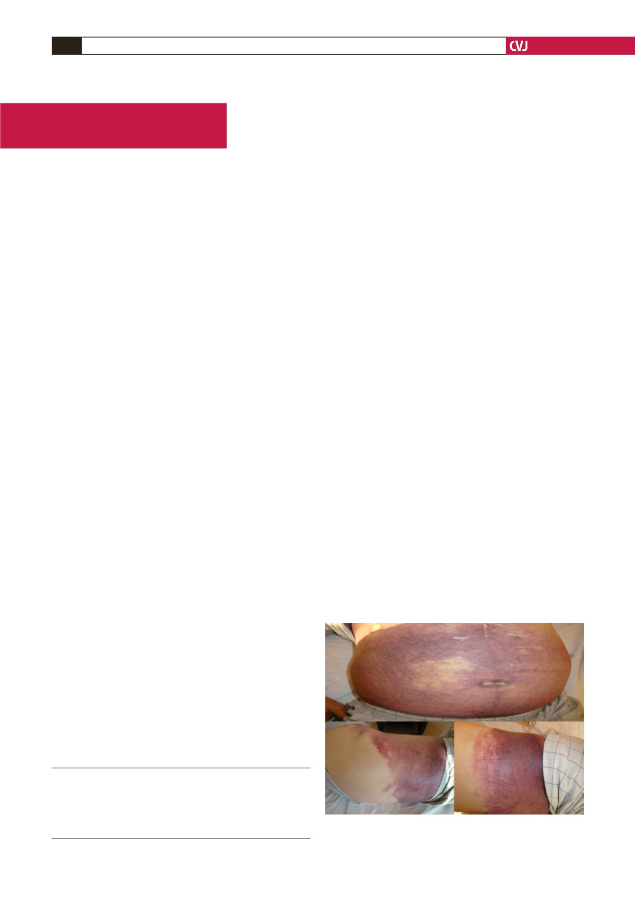
Physical examination revealed mild abdominal swelling,
ecchymosis of the abdomen (positive Cullen’s sign) radiating
to both flanks (positive Gray Turner’s sign) and a palpable mass
on both sides of the umbilicus (Fig. 1). He had tenderness and
mild guarding in the lower quadrants with positive Fothergill’s
and Carnett’s signs. The bowel sounds were normal. He had
tachycardia (120 beats/min) and hypotension (75/40 mmHg).
Blood tests revealed mild leucocytosis accompanied by
anaemia (8.2 g/dl). His INR was 3.0. His abdominal X-ray
was normal and the stool occult blood test was negative.
Transthoracic echocardiography was normal with normal-
functioning prosthetic heart valves and a left ventricular ejection
fraction of 65%. Abdominal ultrasonography showed a large
right-sided RSH, 12 × 22 cm in size.
The patient was transferred to the intensive care unit and
1 mg intravenous vitamin K and three units of fresh frozen
plasma were administered to reverse anticoagulation. Meanwhile
his current medication was immediately stopped and he was
resuscitated with crystaloids and packed red blood cells.
Over the next 12 hours the patient’s symptoms improved
Department of Cardiology, Kartal Kosuyolu Heart Education
and Research Hospital, Istanbul, Turkey
AHMET AYKAN, MD,
ALİ OGUZ, MD
MUSTAFA YİLDİZ, MD, PhD
MEHMET ÖZKAN, MD
Fig. 1. Ecchymosis of the abdomen and flank area is
evident.


