CARDIOVASCULAR JOURNAL OF AFRICA • Vol 23, No 6, July 2012
AFRICA
e17
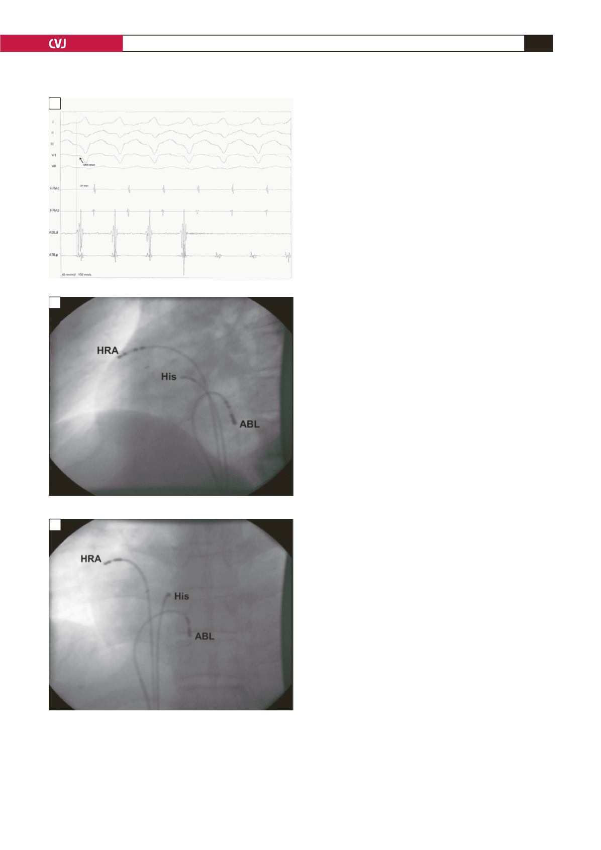
Fig. 2. Intracardiac recordings. A: During the tachycardia,
the ventricular potential recorded by the distal ablation
catheter (ABLd) preceded the onset of the QRS complex
by 27 ms. B: Radiographs obtained in the left anterior
oblique, and C: right anterior oblique projections show
the ablation sites. The distal electrode of the ablation
catheter (ABL) was positioned at the posteroseptal
portion of the tricuspid annulus.
was unremarkable and he had no previous health problems. The
patient’s symptoms started two days earlier and worsened over
time. The palpitations became sustained and he started suffering
from shortness of breath.
The initial 12-lead electrocardiogram (ECG) showed
sustained wide QRS-complex tachycardia consistent with VT
with left bundle branch block (LBBB) with a leftward axis
(Fig. 1). The physical examination was otherwise unremarkable.
Echocardiography revealed a normal left ventricular systolic
function with an ejection fraction of 65%, and no structural heart
disease.
The LBBB pattern and the leftward axis during VT suggested
the inferior wall to be the source of arrhythmia. The patient
underwent an electrophysiological study (EPS). Electro-
anatomical mapping indicated the posteroseptal portion of the
tricuspid annulus as the earliest ventricular activation site during
clinical tachycardia. The intracardiac electrocardiographic
recordings at that region demonstrated a distinct, sharp ventricular
activation 27 ms before the onset of QRS (Fig. 2).
After delivering several RF energy applications (70°C, 50 W)
to the earliest ventricular activation site, the tachycardia suddenly
ceased (Fig. 3). No spontaneous VT/PVCs were observed after
the procedure, and provocation with isoproterenol failed to
induce any tachycardia after the procedure.
His surface ECGs were completely normal in the two
days before discharge and the patient was discharged from
hospital without anti-arrhythmic medications. During the
subsequent six-month follow up, he remained asymptomatic
and no ventricular ectopic activity originating from the posterior
tricuspid annulus was deteceted in 24-hour Holter recordings.
Discussion
Idiopathic right ventricular tachycardia mostly originates from
the septal region of the RVOT, and the ECG shows LBBB
rightward axis morphology. Idiopathic VT/PVCs in the absence
of any underlying structural disease are considered to be
benign.
1
However, occasionally VT/PVCs originating from the
right ventricle may be the first and only manifestation of
arrhythmogenic right ventricular dysplasia, often a familial
disease that can cause sudden death.
Tricuspid annular VT is a rarely encountered entity. Tada
et al
.
2
reported that 8% of idiopathic VT/PVCs originate from
the tricuspid annulus. Their well-designed study demonstrated
that the anteroseptal annulus was the most common site to host
arrhythmogenic foci around the tricuspid annulus.
2
They reported
that ECG findings were useful in identifying the precise origin
of the VT/PVCs arising from the tricuspid annulus.
2
In our case,
LBBB morphology accompanied with leftward axis deviation on
the surface ECG suggested that the arrhythmogenic focus was
the posteroseptal tricuspid annulus.
Tada
et al
.
2
demonstrated that VT/PVCs originating from
the tricuspid annulus had LBBB block, QRS morphology
and positive QRS polarity in leads I, V5 and V6. No negative
deflection was observed in lead I. Since VT/PVCs originating
from the tricuspid annulus were initiated from the right anterior
wall of the heart, an rS or QS pattern in lead aVR was observed
on the surface ECG, similar to RVOT VT/PVCs. However, in
lead aVL, a QS or rS pattern was rare (8%), and the QRS polarity
in lead aVL was positive in almost all VT/PVCs originating from
A
B
C


