CARDIOVASCULAR JOURNAL OF AFRICA • Vol 23, No 6, July 2012
e16
AFRICA
Case Report
Sustained idiopathic ventricular tachycardia originating
from the posteroseptal tricuspid annulus
TURGAY CELIK, BARİS BUGAN, SEDAT KOSE, U CAGDAS YUKSEL, ATİLA IYISOY, H KUTSİ KABUL,
HURKAN KURSAKLIOGLU, ERSOY ISIK
Abstract
Tricuspid annular ventricular tachycardia (VT) is a rarely
encountered entity. Despite abundant data on idiopathicVTs,
the prevalence and clinical characteristics of this infrequent
form are not well defined and the efficacy of radiofrequency
(RF) catheter ablation treatment remains unknown. We
report on a case of a 44-year-old male presenting with symp-
tomatic sustained idiopathic VT originating from the poster-
oseptal tricuspid annulus.
Keywords:
tricuspid annulus, idiopathic ventricular tachycardia
Submitted 16/2/10, accepted 16/2/12
Cardiovasc J Afr
2012;
23
: e16–e18
DOI: 10.5830/CVJA-2012-010
Idiopathic ventricular tachycardias (VT) and premature
ventricular contractions (PVC) mostly originate from the right
ventricular outflow tract (RVOT). In contrast to abundant data on
VTs originating from the RVOT, idiopathic VT/PVCs originating
from the tricuspid annulus have been reported in only a few
cases. Anatomical localisation of arryhthmogenic foci and the
electrophysiological characteristics of this infrequent tachycardia
are obscure and the efficacy of radiofrequency (RF) catheter
ablation of idiopathic VT/PVCs therapy remains unknown.
1
We
report a case of a 44-year-old male presenting with symptomatic
sustained idiopathic VT originating from the posteroseptal
tricuspid annulus.
Case report
A 44-year-old male was admitted to our hospital with palpitations,
dyspnoea on exertion, and a syncopal attack. His medical history
Department of Cardiology, School of Medicine, Gulhane
Military Medical Academy, Etlik, Ankara, Turkey
TURGAY CELIK, MD,
SEDAT KOSE, MD
U CAGDAS YUKSEL, MD
ATİLA IYISOY, MD
H KUTSİ KABUL, MD
HURKAN KURSAKLIOGLU, MD
ERSOY ISIK, MD
Department of Cardiology, Malatya Army District Hospital,
Malatya, Turkey
BARİS BUGAN, MD
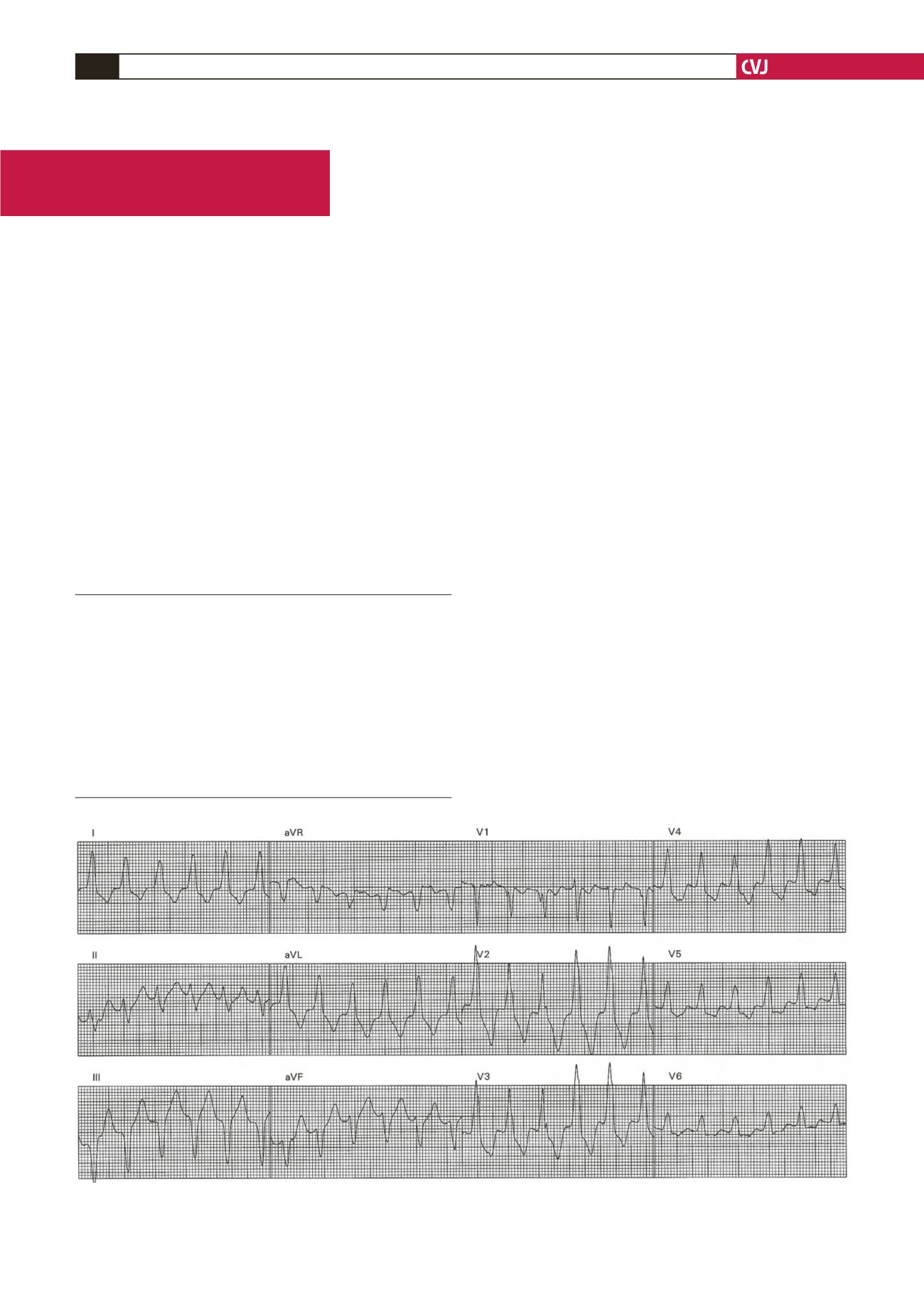
Fig. 1. A 12-lead ECG showing the sustained ventricular tachycardia with QRS morphology of left bundle branch block
with a leftward axis.


