


 71 / 78

CARDIOVASCULAR JOURNAL OF AFRICA • Volume 30, No 1, January/February 2019
AFRICA
e1
Case Report
Delayed left ventricular pseudo-aneurysm after post-
infarction repair of ventricular septal defect
Yun-Seok Song, Sang-Hoon Seol, Seunghwan Kim, Dong-Kie Kim, Ki-Hun Kim, Doo-Il Kim,
Hee-Jae Jun
Abstract
Left ventricular pseudo-aneurysm is a rare complication that
usually occurs after myocardial infarction or cardiac surgery.
Sometimes it is related to cardiac rupture. We report on surgi-
cal management for a left ventricular pseudo-aneurysm that
developed four years after surgery for ventricular septal defect
in a patient with acute myocardial infarction.
Keywords:
pseudo-aneurysm, ventricular septal defect, myocar-
dial infarction
Submitted 20/12/17, accepted 1/10/18
Published online 6/11/18
Cardiovasc J Afr
2019;
30
: e1–e3
www.cvja.co.zaDOI: 10.5830/CVJA-2018-049
Left ventricular (LV) pseudo-aneurysm or false aneurysm is
a free-wall rupture contained by pericardial adhesion or the
epicardial wall. Although the most common cause is myocardial
infarction (MI), it can occur after heart surgery, trauma or
infection.
1,2
We report on a case of a 77-year-old woman with LV
pseudo-aneurysm after post-infarction repair of a ventricular
septal defect (VSD).
Case report
A 77-year-old woman with a history of post-infarction repair of
VSD was noted on regular check-up echocardiography to have
an extra-cardiac mass. She had no symptoms. Four years earlier,
she had presented with acute anterior wall MI with VSD and
Division of Cardiology, Inje University College of Medicine,
Haeundae Paik Hospital, Busan, Korea
Yun-Seok Song, MD
Sang-Hoon Seol, MD,
shseol@paik.ac.krSeunghwan Kim, MD
Dong-Kie Kim, MD
Ki-Hun Kim, MD
Doo-Il Kim, MD
Division of Thoracic Surgery, Inje University College of
Medicine, Haeundae Paik Hospital, Busan, Korea
Hee-Jae Jun, MD
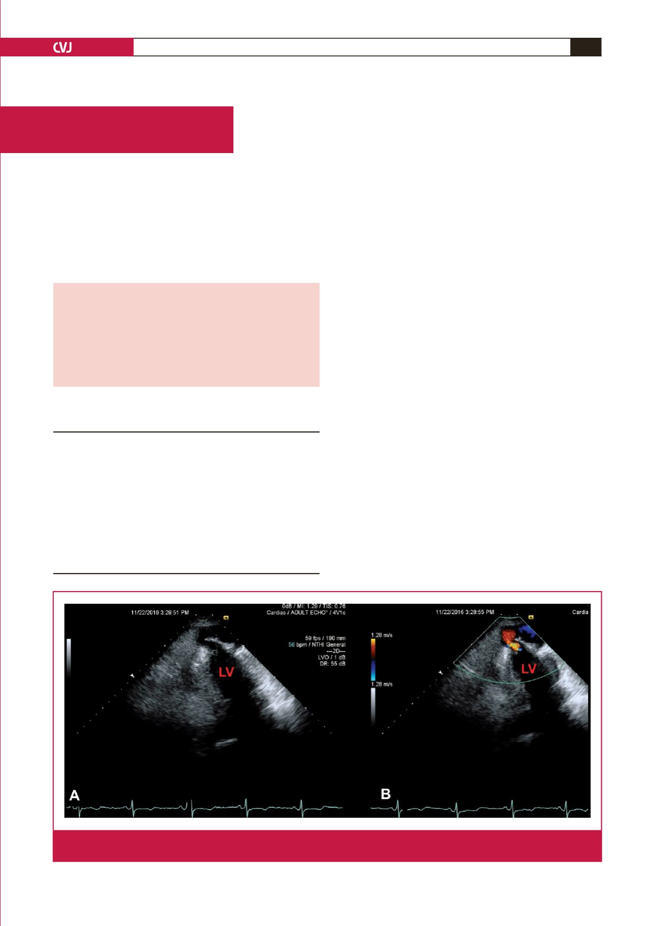
Fig. 1.
Transthoracic echocardiography in the apical two-chamber view reveals a large extra-cardiac echo-free space adjacent to
the LV apex (A), with colour Doppler image between the two (B). LV, left ventricle.